
The International Tinnitus Journal
Official Journal of the Neurootological and Equilibriometric Society
Official Journal of the Brazil Federal District Otorhinolaryngologist Society

 44 7897 074717
ISSN: 0946-5448

Google scholar citation report
Citations : 12717
The International Tinnitus Journal received 12717 citations as per google scholar report

The International Tinnitus Journal peer review process verified at publons

Indexed In
- Excerpta Medica
- Scimago
- SCOPUS
- Publons
- EMBASE
- Google Scholar
- Euro Pub
- CAS Source Index (CASSI)
- Index Medicus
- Medline
- PubMed
- UGC
- EBSCO
Volume 30, Issue 1 / June 2026
Research Article Pages:8-13
10.5935/0946-5448.2026002
A family with audible and non-audible tinnitus revealed by spontaneous otoacoustic emission recordings
Authors:
Nodoka Adachi,Satoshi Asanuma,Naoko Imai,Toshiko Kamishima,Shinsaku Matsuda,Kimitaka Kaga,
Abstract
Objective: Spontaneous otoacoustic emissions (SOAEs) are continuous signals that are generated in the cochlea despite the absence of external auditory stimulation. We studied SOAEs in a male infant with persistent and highfrequency audible tinnitus arising from the inner ear and in his family members (his parents and elder sisters) who have no symptoms. Method: SOAE, distortion product otoacoustic emission (DPOAE), and auditory brainstem response (ABR) tests were conducted to objectively evaluate auditory functions. The patient was a one-year-old male infant with audible tinnitus and his parents and two elder sisters had non-audible tinnitus. For the last three years, their SOAEs were repeatedly recorded. Results: Seven SOAE tests performed over three years demonstrated positive peaks with the main peak at 5.3 kHz on the left side of the infant. His 42-year-old father, 5-year-old sister, and 7-year-old sister without objective tinnitus showed SOAEs, but his 38-year-old mother did not show any SOAEs. Conclusion: The results of frequent SOAE tests over three years suggested that the audible tinnitus in the infant and the non-audible tinnitus in the family members could have arisen from the spontaneous vibration of outer hair cells. However, a particular energy mechanism can cause the vibration of outer hair cells to produce audible SOAEs. Our study suggests the possibility of a familial inheritance of SOAE in this family.Keywords:
SOAE, audible tinnitus, outer cell, inheritance.
Introduction
Spontaneous otoacoustic emissions (SOAEs) are continuous signals that are generated in the cochlea despite the absence of external auditory stimulation [1]. They occur in more than half of normal ears, including those of infants, children, and adults. Typically, SOAEs measured in adults are concentrated in the frequency range of 1.0 to 2.0 kHz and have been observed at frequencies from 0.5 to 9.0 kHz. In general, SOAEs are not seen in frequency regions in patients with sensorineural hearing loss of lower than 30 dB HL. It is considered that SOAE originates from outer hair cells corresponding to the portion of the basilar membrane tuned to that frequency [2]. Before OAE recordings became available, four possible origins were theorized by Glanville et al. in 1971 [3]: (1) resonance, (2) middle ear muscle fasciculation, (3) reverse neuroacoustic transduction from the internal ear, and (4) vascular sound.
There are persons who emit audible sounds that can be heard by other people. Such sounds are called SOAEs. Their origin, which has not been precisely demonstrated, appears to be the outer hair cells of the cochlea. SOAEs are classified into low-intensity non-audible SOAE and high-intensity audible SOAE [4]. In this study, we investigated SOAEs in a male infant with persistent and high-frequency audible tinnitus probably arising from the inner ear and in his family members (his parents and two elder sisters) without audible tinnitus.
Materials and Methods
Subject
The patient was a male infant aged 1 year at the time of the initial visit. He was born by normal delivery at 39 weeks of gestation and weighed 2308 g. After birth, he was hospitalized with hypoglycemia in a neonatal intensive care unit for three days. The outcome of the neonatal automated auditory brainstem response (AABR) screening showed “pass” in both ears. No external malformation was observed. At six months, his mother noticed a high-frequency sound from the right ear. At eight months, he was brought to a local otolaryngologist because of nasal discharge and was diagnosed as having secretory otitis media in both ears. The high-frequency sound from the right ear temporarily disappeared at that time. At nine months, the high-frequency sound was again noticed as the otitis media in both ears improved, but at this time, it came from the left ear.
At one year old, he and his family were referred to our Children’s Medical Center by a local otolaryngologist for further examinations. The high-frequency sound from the left ear persisted and was audible from a distance of about 30 cm in a soundproof room in the outpatient clinic. In our observation for the last three years, the sound persisted at a constant high frequency and intensity and did not change at all.
Family Tree
Figure 1 RPatient’s family tree.
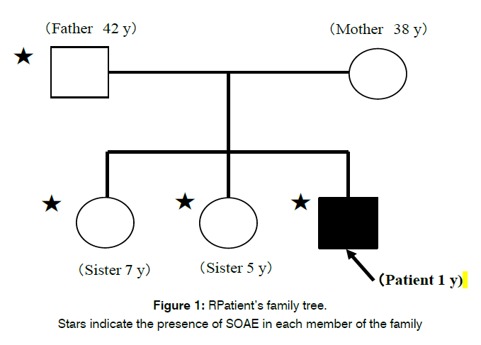
Figure 1: RPatient’s family tree.
Stars indicate the presence of SOAE in each member of the family
Methods
The SOAE test was performed using ILO292-USB ver.6 (RION). Objective audiometry was performed using ABR test and behavioral observation audiometry was performed using the conditioned orientation reflex (COR) test. Imaging studies of the middle ear and inner ear were conducted by computed tomography (CT) and magnetic resonance imaging (MRI).
Results
Patient’s study results
Behavioral and objective audiometry
The hearing threshold level determined by COR audiometry was normal at 30 dB nHL. ABR showed a normal wave Ⅴ threshold at 20 dB nHL in both ears of the patient.
Imaging studies
The CT and MRI scans of the temporal bone demonstrated normal middle ear and inner ear on both sides.
SOAE study
Figure 2 shows SOAE recordings at four different over the last three years.
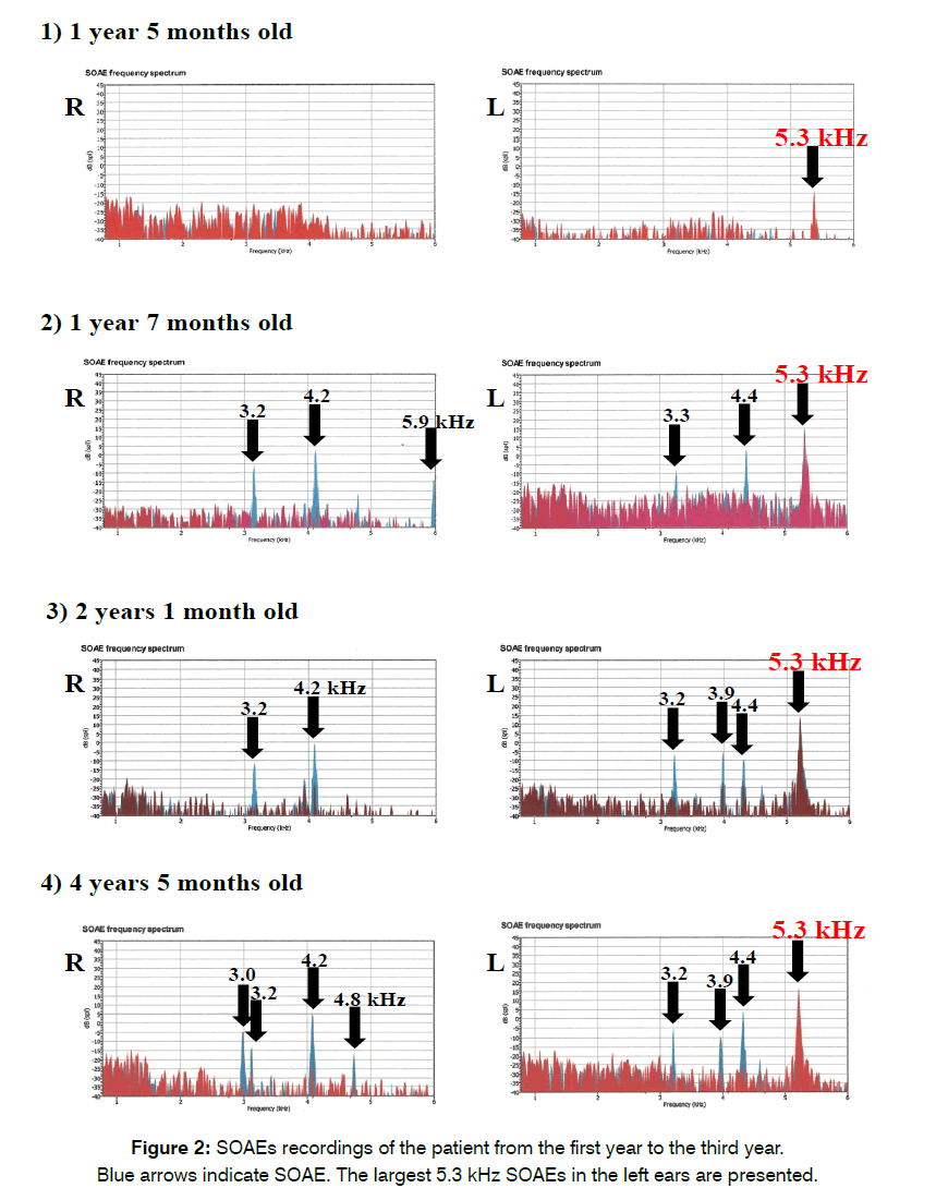
Figure 2: SOAEs recordings of the patient from the first year to the third year.
Blue arrows indicate SOAE. The largest 5.3 kHz SOAEs in the left ears are presented.
1. At the age of one year and five months, there was a peak at 5.3 kHz (− 14 dB SPL) in the left ear but none in the right ear.
2. At the age of one year and seven months, there were peaks at 3.2 kHz (− 6 dB SPL), 4.2 kHz (+ 3 dB SPL), and 5.9 kHz (− 14 dB SPL) in the right ear, and at 3. 3 kHz (− 9 dB SPL), 4.4 kHz (+ 3 dB SPL), and 5.3 kHz (+ 15 dB SPL) in the left ear.
3. At the age of two years and one month, there were peaks at 3.2 kHz (− 11 dB SPL) and 4.2 kHz (− 1 dB SPL) in the right ear and at 3.2 kHz (− 7 dB SPL), 3.9 kHz (− 6 dB SPL), 4.4 kHz (− 10 dB SPL), and 5.3 kHz (+ 14 dB SPL) in the left ear.
4. At age of four years and five months, there were peaks at 3.0 kHz (− 5 dB SPL), 3.2 kHz (− 14 dB SPL), 4.2 kHz (+ 5 dB SPL), and 4.8 kHz (− 17 dB SPL) in the right ear, and at 3.2 kHz (− 6 dB SPL), 3.9 kHz (− 10 dB SPL), 4.4 kHz (− 10 dB SPL), 5.3 kHz (+ 16 dB SPL) in the left ear.
Family members’ study results
Four other family members (father, aged 42; mother, aged 38; 5-year-old sister, 7-year-old sister) underwent SOAE tests.
Figure 3 shows SOAE recordings of his father and two elder sisters.
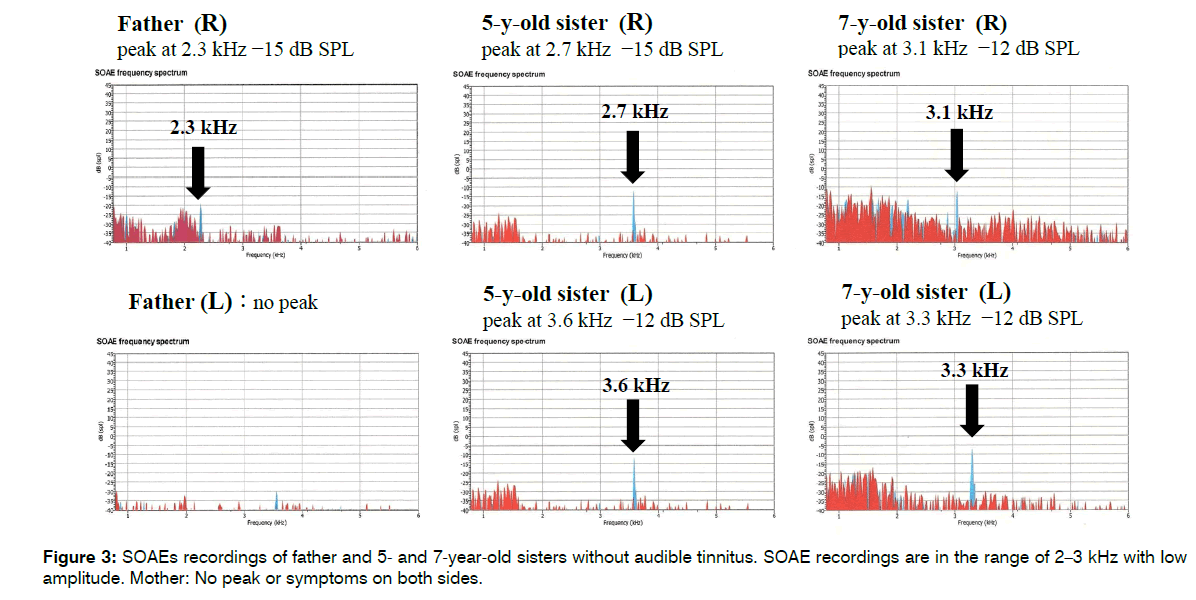
Figure 3: SOAEs recordings of father and 5- and 7-year-old sisters without audible tinnitus. SOAE recordings are in the range of 2–3 kHz with low amplitude. Mother: No peak or symptoms on both sides.
His father had a peak at 2.3 kHz (− 20 dB SP) in the right ear, which was smaller than that of the patient, with normal findings in the left ear. His 5-year-old sister had small peak at 2.7 kHz (−14 dB SPL) in the right ear and 3.6 kHz (− 12 dB SPL) in the left ear. His 7-year-old sister had small peak at 3.1 kHz (− 13 dB SPL) in the right ear and 3.3 kHz (− 13 dB SPL) in the left ear. On the other hand, no SOAE peak was observed in either ear of his mother.
Discussion
Kaga et al. reported the following features of high-intensity audible SOAEs in children: (1) OAEs with levels up to 60 dB SPL are rare; (2) audible without amplification; (3) high-frequencies; (4) present at birth; (5) may be inherited; (6) a particularly sensitive cochlea; (7) hearing is always normal even when SOAE is present [4].
In our study, only the male infant manifested audible tinnitus (SOAE), whereas his family members did not show any audible tinnitus. However, SOAE tests demonstrated similar SOAE peaks among the male infant, in his father, and two older sisters. The recorded SOAEs can show the probability of inheritance of SOAE, as a familial phenomenon except for his mother. This audible tinnitus is different from objective tinnitus that is characterized by discontinuous and pulsatile clicks caused by the periodic contraction of pharyngeal muscles caused by pharyngeal myoclonus. We have followed up the patient and his family members over three years to determine whether the congenital SOAE is a temporary phenomenon or not. The patient’s audible tinnitus and SOAE continuously appeared over the last three years.
Glanville et al. [3] reported three cases of a father and two of his three children with high-frequency non-pulsatile audible sounds emitted at different pure tones from five of six ears in this family. The condition is considered inherited as a dominant characteristic that is not sex-linked. Table 1 shows the frequency analysis of SOAEs in children in the literature (congenital type).
| Source, year | Patient’s age/sex | Affected ear | Tone | |
|---|---|---|---|---|
| Frequency(Hz) | Level(dB SPL) | |||
| Labell, 1962 | 3.5 years/M | Both | R:not mentioned | Not mentioned |
| L:5020 | ||||
| Citron, 1969 | 4 years/F | Both | R:8000 | Not mentioned |
| L:not mentioned | ||||
| Glanville et al., 1971 | 4 years/F | Both | R:7630 | R: 35 |
| L:5640 | L: 38 | |||
| Glanville et al., 1971 | 3.6 years/M | Both | R:8470 | R: 21 |
| L:8410 | L: 24 | |||
| Mathis et al., 1991 | 6 months/M | Left | L:5640 | L: 55 |
| Kaga et al., 2009 | 5 months/F | Both | R:4200, 6500 | Both: 42 |
| L:2400 | ||||
| Kaga et al., 2009 | 11 months/F | Left | L:6700 | L: 42 |
| Present patient | 1 year/M | Both | 5300 | L : 14 |
Table 1: Frequency analysis of SOAEs in children (congenital type) in the literature [5].
Two types of SOAE can be identified [5, 6]: (1) the common low-intensity non-audible type that can be recorded in the majority of normal-hearing ears and (2) the rare high-intensity audible type that is clearly related to the pathological hearing of children [3, 5, 7-15] as shown in Table 1 and adults as shown in Table 2 [3, 12, 13].
| Source, year | Patient’s age/sex | Affected ear | Tone | |
|---|---|---|---|---|
| Frequency(Hz) | Level (dB SPL) | |||
| Glanville et al., 1971 | 26 years/M | Left | L: 6230 | L: 20 |
| Huizing and Spoor, 1973 | 22 years/F | Right | R: 3400 | R: 35 |
| Yamamoto et al., 1987 | 25 years /M | Right | R: 6100 | L: 37.2 |
Table 2: Frequency analysis of SOAEs in adults (acquired type) in the literature [5].
The features of low-intensity non-audible SOAEs are as follows: (1) found in up to 78% of subjects with presumed normal hearing; (2) a byproduct of normal cochlear functioning; (3) normal audiometric thresholds; (4) a typical level of about 1 dB SPL ranging from − 10 to + 20 dB SPL in adults; (5) relatively high-frequency [5]. In the literature, SOAEs are classified into the congenital type (Table 1) and the acquired type (Table 2). The SOAEs in the affected family members of this study were of the congenital high-intensity audible type, and probably, SOAEs at a high-frequency may be caused by a spontaneous and continuous contraction of outer hair cells, resulting in the emission of audible sounds from their ears. If this theory were true, outer hair cells would lose their inhibitory function or augment active functions at a high frequency. The SOAE in our patient has continued up to his present age of four years.
Regarding the mechanism of OAE, it was first postulated by Gold in 1948 that the ‘cochlea does not only receive sounds but can also produce acoustic energy.’ [12]. From acoustic impulse response of human ear recordings, Kemp discovered the ’cochlea echo’ as OAE in 1978 [13]. Transient evoked otoacoustic emission (TEOAE) or DPOAE is evoked by otoacoustic emissions as an electrical phenomenon of cochlear origin in response to the input sound. However, spontaneous emission can be practically recorded without any input sounds to study ‘the cochlear origin of tinnitus’. Table 3 shows the list of hypotheses [2, 5, 13]. The number of cases of objective tinnitus caused by inner ear problems is extremely small. There are only a few hypotheses presented that clarified the cochlear origin of objective tinnitus. Kemp suggested there is a biological–mechanical mechanism in the cochlea that amplifies a sound wave, which could be related to TEOAE, DPOAE, and SOAE [13].
| Author name | Year | Hypotheses |
|---|---|---|
| Kemp | 1978 | An active wave amplification mechanism in the cochlear is proposed. |
| Zurek | 1981 | The factors for active biomechanical processes are present in the inner ear and support the processes that increase the sensitivity and selectivity of the mechanical frequency analysis of the cochlea. |
| Kaga | 2009 | It may be caused by the spontaneous and continuous contraction of outer hair cells, resulting in the emission of audible sounds from their years. |
Table 3. List of hypotheses on audible tinnitus.
Zurek suggested that the factors for active biomechanical processes are present in the inner ear and support the processes that increase the sensitivity and selectivity for the mechanical frequency in the cochlea [2].
Conclusion
The results of frequent SOAE tests over three years suggest that the audible tinnitus in our patient and could have arisen from the spontaneous vibration of outer hair cells and the non-audible tinnitus in his family members. Probably, an energy mechanism can cause the vibration, which can produce audible SOAEs. We regarded SOAE related to audible tinnitus as a unique phenomenon caused by spontaneous outer hair cell hyperactivity.
The results of our study, there is a possibility of a familial inheritance.
Acknowledgments
We thank Ms. Chiemi Shoji and Ms. Kayoko Sekiguchi for their secretarial assistances.
Declaration of Conflict of Interest
The authors report no conflicts of interest. The authors are responsible for the content and writing of the paper.
References
- Prieve B, Fitzgerald T. Otoacoustic emissions. Handbook of clinical audiology. 2002;5:440-66.
- Zurek PM. Spontaneous narrowband acoustic signals emitted by human ears. J Acoust Soc Am. 1981;69(2):514-23.
- Glanville JD, Coles RR, Sullivan BM. A family with high-tone objective tinnitus1. J Laryngol Otol. 1971;85(1):1-0.
- HARRIS FP. Otoacoustic emissions and audiometric outcomes. OAE. 1997:151-80.
- Kaga K, Akamatsu Y, Nakamura M, Sakata H. Spontaneous otoacoustic emissions in two infants. Acta Oto-Laryngol. 2009;129(4):399-404.
- Probst R, Lonsbury-Martin BL, Martin GK. A review of otoacoustic emissions. J Acoust Soc Am. 1991;89(5):2027-67.
- Loebell E. Demonstration of a patient. HNO. 1962;10:223-4.
- Citron L. Observations on a case of objective tinnitus. Int Congr Ser. 1969;189:91.
- Mathis A, Probst R, De Min N, Hauser R. A child with an unusually high-level spontaneous otoacoustic emission. Arch. Otolaryngol. Head Neck Surg. 1991;117(6):674-6.
- Sato N, Toshitsuru H, Sakai N, Terayama Y, A family case of spontaneous otoacoustic emission, Clin Otol Jpn. 14 (1987) 208
- Suzuki M, Tanaka Y, Inoue T, Tuzuku T. An infant with audible spontaneous otoacoustic emissiom. Audiol Jpn. 1990;33(2):164-8.
- Huizing EH, Spoor A. An Unusual Type of Tinnitus: Production of a High Tone by the Ear. Arch Otolaryngol. 1973;98(2):134-6.
- Yamamoto E, Takagi A, Hirono Y, Yagi N. A Case of'Spontaneous Otoacoustic Emission'. Arch Otolaryngol Head Neck Surg. 1987;113(12):1316-8.
- Gold T. Hearing. II. The physical basis of the action of the cochlea. Proc. R. Soc. B: Biol. Sci. 1948;135(881):492-8.
- Kemp DT. Stimulated acoustic emissions from within the human auditory system. J Acoust Soc Am. 1978;64(5):1386-91.
1Division of Otolaryngology, Saitama Children’s Medical Center, 1-2 Chuo-ku, Saitama-shi, Saitama, 330-8777, Japan
2Department of Clinical Laboratory, Saitama Children’s Medical Center, 1-2 Chuo-ku, Saitama-shi, Saitama, 330-8777, Japan
3Department of Otorhinolaryngology and Neck and Head Surgery, University of Tokyo, 7-3-1 Hongo, Bunkyo-ku, Tokyo, 113-8655, Japan
4National Institute of Sensory Organs, NHO Tokyo Medical Center, 2-5-1, Higashigaoka, Meguro-Ku, Tokyo, 152-8902, Japan
5Kamio Memorial Hospital, 2-25 Kanda-Awajicho, Chiyoda-ku, 101-0063, Tokyo, Japan
Send correspondence to:
Nodoka Adachi, M.D
Saitama Children’s Medical Center, 1-2 Chuou-ku, Saitama-shi, Saitama, Japan. E-mail : nodoka05270527@gmail.com
Tel : +81 48 601 2200
Paper submitted on May 30, 2026; and Accepted on June 03, 2026
Citation: Nodoka A. A Family with Audible and Non-Audible Tinnitus Revealed by Spontaneous Otoacoustic Emission Recordings. Int Tinnitus J. 2026;30(1): 08-13.