The International Tinnitus Journal
Official Journal of the Neurootological and Equilibriometric Society
Official Journal of the Brazil Federal District Otorhinolaryngologist Society
ISSN: 0946-5448

Google scholar citation report
Citations : 12717
The International Tinnitus Journal received 12717 citations as per google scholar report
The International Tinnitus Journal peer review process verified at publons
Indexed In
- Excerpta Medica
- Scimago
- SCOPUS
- Publons
- EMBASE
- Google Scholar
- Euro Pub
- CAS Source Index (CASSI)
- Index Medicus
- Medline
- PubMed
- UGC
- EBSCO
Volume 24, Issue 2 / December 2020
Research Article Pages:79-85
10.5935/0946-5448.20200016
Association between Spontaneous Otoacoustic Emission and Psychoacoustic measures
Authors: Kaushalendra Kumar, Jobby John, Rohit Ravi
PDF
Abstract
Aim: The aim of the present study was to evaluate the association of presence and absence of spontaneous otoacoustic emissions (SOAEs) on different psycho-acoustic measures such as intensity discrimination, gap detection test, duration discrimination test, modulation detection for sinusoidal amplitude modulated noise at 8, 20, 60, and 100 Hz.
Method: Sixty adults with hearing sensitivity within normal limits were divided into two groups; group 1 consisted of participants with SOAEs present and group 2 consisted of participants with SOAEs absent. All the participants were tested for presence of SOAEs and different psycho-acoustic measures.
Results: The present study results showed no significant difference on intensity discrimination, gap detection test, duration discrimination test, modulation detection for sinusoidal amplitude modulated noise at 8, 20, 60, and 100 Hz in presence and absent of SOAE.
Conclusion: The findings reveals that the presence or absence of SOAE did not influence or enhance the psychophysical performance at most comfortable level in individuals having normal hearing. Keywords: Psycho-acoustic measures, spontaneous otoacoustic emissions, gap detection
Keywords: Psycho-acoustic measures, spontaneous otoacoustic emissions, gap detection
Introduction
Otoacoustic emissions (OAEs) simple, efficient and noninvasive and an objective indication of healthy cochlear function [1]. OAEs are sounds which arise in the ear canal when the tympanum receives vibrations that are transmitted backwards from the cochlea through the middle [2]. Spontaneous OAEs (SOAEs) occur without any external acoustic stimulation and consist of energy at one or more frequencies emitted by the normal ear. SOAEs are emitted as a result from the autonomous mechanical oscillation of cellular or sub cellular constituents of the ear’s amplifier [3]. SOAEs are detected with a sensitive and low noise microphone housed within a probe assembly, which is fit snugly into the ear canal. Improved signal to noise levels and minimal noise helps in better detection of SOAEs [4]. Studies indicate that multiple SOAEs are observed in female subjects than male subjects, in ears with more than one SOAE, the minimum frequency difference is about 100 Hz and is rarely less than 50 Hz [5-7]. Talmadge et al. 8 have suggested that the minimum separation between SOAEs corresponds to about one twelfth octave (or a distance of about 0.4 mm on the basilar membrane).
Psychoacoustic tests: Psychoacoustic tests involve a subjective evaluation of the how the sound heard is perceived by a person. Gap detection test is one of the psycho acoustical tests, which is well researched and assess for the temporal resolution of auditory system. The listeners are required to detect a brief pause in an otherwise continues sound. Central auditory processing is a common term that is being used among audiologists and speech language pathologists. These processing is responsible for localization and lateralization of sound, auditory discrimination and even temporal aspect of audition (temporal resolution, temporal masking [8], temporal integration and temporal ordering); both verbal and non-verbal signals are processed and impairment may affect areas with function of speech and language. The central auditory processing disorder can be confirmed using GIN (Gap-In-Noise) test [9]. Since GIN test and RGD test are sensitive to central auditory nervous system lesion, both these tests are used as a best tool to assess in clinical population with CAPD
Duration discrimination is a temporal task where a reference signal at a fixed duration should be discriminated from a comparison signal, which has duration different from the reference signal. The reference signal can be tonal or noise. Since duration discrimination is more of perceptual level, it can be affected by age, where discrimination of the brief temporal gap (6.4 msec) was influenced by age and hearing loss, i.e., duration discrimination scores were reduced in elderly population for both tones and silent intervals when the reference duration was 250 msec [10]. Another psychophysical test procedure which checks the minimum intensity to identify as two different sounds accounts for the temporal resolution of audit, here the subject’s ability to differentiate the minimum difference in intensity (dB) is estimated. The intensity discrimination task is a complex process which involves some amount of physiological changes and is influenced by temporal aspect [11].Temporal modulation detection is a test done to examine the temporal resolution where a minimal amount of Sinusoidal Amplitude Modulated signal is presented and listener need to discriminate between modulated and unmodulated noise. Temporal modulation transfer function is a result of modulation thresholds as a function of frequency of modulation. Modulation thresholds are expressed in dB and are calculated as 20 log m, where m is index of modulation.
Oto-acoustic emissions and psycho-acoustics
Few studies have been accounted for the influence of otoacoustic emissions on the peripheral auditory processing. Smurzynski et al. [12] studied influence of Spontaneous otoacoustic emissions on gap detection threshold. Results showed both the groups significantly differed in 10 dB SL conditions where the group I, with present SOAE, confirmed a better gap detection threshold and outperformed group II having absent SOAE. They explained this as near the hearing threshold a short gap is masked by SOAEs and OAEs that are evoked by the stimulus. This effect is stronger for ears with multiple and robust SOAEs. For higher stimulus levels, SOAEs are suppressed by the test signal. The gap detection threshold is shorter than the time needed to recover from suppression. Smurzynski et al. [12] measured intensity discrimination, temporal integration, and gap detection in normal hearing individuals with strong and weak OAEs and found that OAEs can influence performance on these psychoacoustical tasks, especially for low-level stimuli with spectral components in the vicinity of high-level SOAEs.
Need for the study
Previous studies have indicated that a better gap detection threshold in subjects with present SOAE at low level SL, however; at higher level there was no effect seen on it. Literature also reports that there is no difference in intensity discrimination task and temporal integration function with SOAE present. However, there is lack of research on effect of present SOAE and Temporal Modulation transfer function test and duration discrimination test. The aim of the present study was to evaluate the association of presence and absence of SOAE on different psycho-acoustic measures. The specific objectives were to; determine the association between SOAE and intensity discrimination, association between SOAE and gap detection in noise, association between SOAE and duration discrimination, association between SOAE and modulation detection for sinusoidal amplitude modulated noise at 8, 20, 60, and 200 Hz.
Methods
This study commenced after clearance from the Institutional ethical board. Informed consent was taken from all the participants after explaining them about the study.
Participants and testing details
Sixty adults with hearing sensitivity within normal limits in age range of 20-40 years participated in the study. They were divided into two groups; group 1 consisted of participants with SOAEs present and group 2 consisted of participants with SOAEs absent. All participants had pure tone thresholds, less than or equal to 15 dB HL in the octave frequencies from 250 Hz to 8 kHz for air conduction and from 250 Hz to 4kHz for bone conduction. All participants had ‘A’ type tympanogram and the acoustic reflexes were present at 500 and 1 kHz at normal sensation levels. A structured case history was taken to confirm that none of the participants had any history of otological or gross neurological deficits, occupational noise exposure and ototoxicity. The testing comprised of measuring SOAEs followed by psychoacoustical tests. The psychoacoustical measures such as intensity discrimination, duration discrimination, gap detection threshold test and temporal modulation test were conducted. For the first three tests, 30 ears with present and absent SOAE were selected while for the last test 15 participants with bilateral SAOE present and absent were selected.
Convenient sampling was used to recruit the participants in each group. The sample size was estimated based on the following The total number of 30 subjects for each group was selected based on convenient sampling with 95 percent confidence interval equal to 1.96 as Zα and 80 percent power a sample equal to 0.84 as Zβ based on the formula,

Where, Zα/2 =value at a specified significance level
Zβ =value at specified type2 error or power
S =pooled standard deviation of observational of the two sample
d= clinical significant difference
Test environment: All the evaluations were carried out in an acoustically treated room with adequate illumination and permissible background noise. Pure tone audiometry was carried out in double- room suite whereas tympanometry and SOAE measurements were done in a single-room suite.
Procedure: Initially, a detailed case history was taken to ascertain the inclusion of the participant. This was followed by hearing evaluation in a sound treated room using a duly calibrated GSI-61 clinical audiometer coupled with TDH-49P headphones to obtain air conduction thresholds. Bone conduction thresholds were obtained using Radio ear B-71 bone vibrator. The participants with pure tone average of 15dBHL or less were selected for the study based on Modified Hughson- Westlake procedure [13]. Middle ear evaluation was done using a calibrated immitance audiometer, GSI-Tympstar Middle Ear Analyzer; with a probe tone of 226Hz to measure the middle ear thresholds. Subjects were instructed not to swallow and move during the test procedure. Participants with bilateral ‘A’ type tympanogram with normal reflexes were selected for the study.
Experimental Tasks
The experimental task consisted of physiological and psycho-acoustic measures. The physiological measure included SOAEs while the psycho-acoustic measures included intensity discrimination, duration discrimination, gap detection threshold test and temporal modulation test.
Physiological experiment- Spontaneous Otoacoustic Emission measurement
Test stimuli and instrumentation: A computer based SOAE analyzer ILO292 was used to record SOAEs. The Spontaneous otoacoustic emission was recorded by coupling a sensitive miniature microphone to the external ear canal. The noise present in the canal was preamplified and filtered using a high pass filter to eliminate physiological noise below 300-500Hz; the obtained signal was delivered to a spectrum analyzer or Fast Fourier transform (FFT) software to do spectral analysis. The SOAEs frequency ranged from 0.6 to 6.9 and majority of SOAEs present in the range of 1 to 3 kHz [14,15] . Subject was instructed to sit back and relax and to reduce his body movements as much as possible. A suitable probe tip was fitted on the probe and inserted into the ear canal of the test ear. Subjects both ear were tested to detect the presence or absence of SOAEs.
Psychoacoustic experiments: The psycho-acoustic tests were done using ‘mlp’ tool box which implements a maximum likelihood procedure in Matlab [16]. Stimuli were recorded at 44,100 Hz sampling rate. The threshold was tracked by two-interval alternate forced choice method using a ‘maximum likelihood procedure’. In every trial, stimuli were presented in each of two intervals: One interval with a reference stimulus, the other interval contained the variable stimulus. After every trial the participant indicated which interval consisted of the variable stimulus. In duration discrimination test, intensity discrimination test and gap detection test the stimuli were presented separately for each ear and binaurally for temporal modulation transfer function test, at comfortable levels. Stimuli were presented via a laptop computer (Hewlett Packard), connected to high fidelity earphones at comfortable level. Subjects were given 3-4 practice trails before the commencement of each test. All psychoacoustic tests were carried out in a quiet room
Duration discrimination test: In this procedure, the minimum difference in duration that was necessary to perceive the two otherwise identical white noise bursts was measured. Duration of the standard stimulus was 250 msec. The task was to tell which interval contained the longer duration signal and the duration of the variable stimulus varied based on participant’s response. Two intervals alternate forced-choice procedure was used to track the threshold.
Gap detection thresholds: The participant’s ability to detect a temporal gap in the center of a 500ms broadband noise was measured. The waveform was digitally shaped at onset and offset with 20ms cosine squared envelopes. The noise had 0.5ms cosine ramps at the beginning and end of the gap. The stimulus presented was 500msec broadband noise with no gap in reference stimulus whereas the variable stimulus contained the gap. A twointerval alternate forced choice procedure was used to track the threshold
Modulation detection thresholds for sinusoidal amplitude-modulated noise: Temporal modulation refers to a reoccurring change (in frequency or amplitude) in a signal over time. A 500msec Gaussian noise was sinusoidal amplitude modulated at modulation frequencies of 8 Hz, 20 Hz, 60 Hz and 200 Hz. Noise stimuli had two 10-msec raised cosine ramps at onset and offset. The participant had to listen to the stimulus and detect the modulation noise. Modulated and un-modulated stimuli were equated for total root mean square (RMS) power. Depth of the modulated signal varied if the participant responded up to an 80% criterion level. The modulation detection thresholds were expressed in dB by using the following equation
Modulation detection thresholds in dB = 20 log10
Data analysis: A present SOAE was a least response at 3dB above noise floor, if not it was considered as absent. Intensity discrimination thresholds and temporal modulation detection thresholds across modulation rates was measured in ‘dB’, while gap detection threshold and duration discrimination threshold was measured in ‘msec’. The thresholds obtained in two blocks were averaged and mean a threshold was obtained.
Statistical analysis: Independent sample t-test was used to compare the findings between the SOAE and the different psychacoustical tests. Statistical package SPSS vers15.0 was used to do the analysis, P< 0.05 was considered as significant.
Results
Intensity discrimination threshold
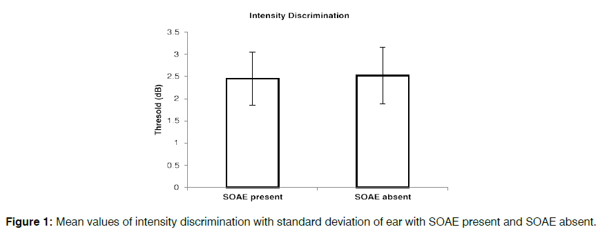
As seen in Figure 1, the mean intensity discrimination threshold was 2.45dB in subject with present SOAE and in absent SOAE it was found to be mean 2.52dB. There was less variation observed in standard deviation. From independent t test it was inferred that there was no significant difference [t (58) = -0.447, P=0.657] in intensity discrimination threshold in subject with present and absent SOAE.
Gap detection threshold and duration discrimination
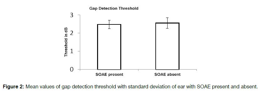
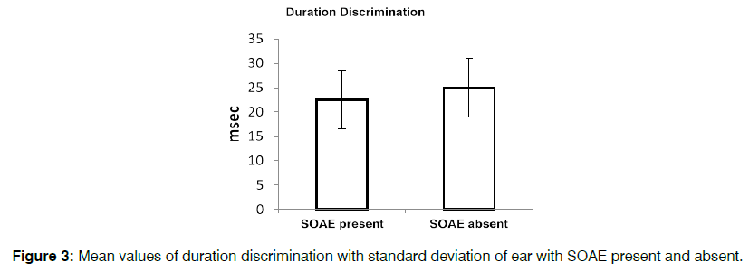
In subjects with present SOAE gap detection threshold mean was observed 2.48msec with standard deviation of 0.24. Similar finding were observed for subject who had absent SOAE (mean 2.56±0.29). Independent t test were performed to see the significant difference in gap detection threshold in subject with present and absent SOAE. From the independent T- test it can be inferred that there was no significant difference [t (58) = -1.21, p=0.657] in gap detection threshold between present and absent SOAE. It can be observed that mean duration discrimination threshold was 22.48 (±5.95) msec with present SOAE and with absent SOAE it was observed to be 25 (±6.02) msec. Independent T-test was carried out to see the significance difference in duration discrimination test in present and absent SOAE. Independent t test revealed that there was no significant difference [t (58) = -1.628, p=0.109] in duration discrimination threshold in present and absent SOAE (Figure 2).
Temporal modulation transfer function
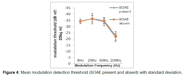
The mean modulation detection threshold at 8Hz, 20Hz, 60Hz, and 200Hz are -34.49, -36.28, -33.80 and -21.38 dB respectively in subject with present SOAE. With present SOAE standard deviation were 0.57, 4.17, 3.09 and 3.29 using 8Hz, 20Hz, 60Hz and 200Hz respectively. In subject with absent SOAE at 8Hz (mean=-43.22±1.86), 20Hz (mean= -37.66±2.67), 60Hz (mean=-34.85±2.30) and 200Hz (mean=-22.47±3.45) Figure 3.It can be observed that as the frequency increases there is decrease in TMTF threshold. Independent t test was performed to see if there is any change in TMTF findings in present and absent SOAE at different modulation frequencies. From the t test it was inferred that there was no significant difference [t(28)=-0.548, p=0.591] in TMTF threshold at 8Hz. Similar findings were observed at 20Hz [t(28)=1.080, p=0.291], 60Hz [t(28)=1.051, p=0.303] and 200Hz [t(28)=-0.890, p=0.381]. Figures 4 shows the mean modulation detection thresholds at different modulation frequencies for ear with SOAE present and ear with SOAE absent respectively. From the figure 4 it is clear that modulation detection thresholds were better for low modulation frequencies and worsened at high modulation frequencies. Hence, the present study suggests there is no effect on TMTF findings in with subject who had either present or absent SOAE.
Discussion
The present study aimed at investigating the association psychophysical measures with the presence and absence of SOAEs. Different psychophysical measures that were considered were intensity discrimination, gap detection in noise, duration discrimination and modulation detection for sinusoidally amplitude-modulated noise at 8, 20, 60, and 200 Hz. In the present study, no significant difference in intensity discrimination threshold in subject with present and absent SOAE was noted. Similarly, Smurzynski et al. [12] had reported no significant difference in intensity discrimination in ear with present and absent SOAE at 60, 40 and 20dB SL. Similar study was done by Probst et al. [17] where they had compared just noticeable difference for intensity in weak and strong OAE at 60, 40 and 20dBSL. Strong OAE were reported as present SOAE and with high level of Transient otoacoustic emission (TEOAE) and weak OAE were reported as absent SOAE with low level TEOAE. Mean just noticeable difference did not significantly vary between two groups weak vs strong OAE. However, intra test variability was observed to be more in subjects with strong OAE as compare to weak OAE. With present and previous study, it can be concluded that SOAE activity does not affect the mean intensity discrimination task.
Gap detection threshold and duration discrimination test
No significant difference was noted in gap detection threshold between present and absent SOAE. Smurzynski et al. [12] performed gap detection test at four different levels as 10, 20, 30 and 50dB SL with present and absent SOAE. They found no significant difference in threshold between present and absent SOAE at 20, 30 and 50dB SL. However; at 10dB SL, there was significant difference in threshold were observed in subject with present and absent SOAE. At lower SL level, threshold was observed to be poorer than higher level in subject with present SOAE is due to close to the audibility threshold suggest that a short gap is masked or partial filled by SOAEs. The effect is stronger for ears with multiple and robust SOAEs. Another hand at higher stimulus levels SOAES are suppressed by the test signal and so both groups performed very similar results as it has been performing in present study. Smurzynski et al. 12 compared temporal integration function in persons with and without SOAE. Subjects with present SOAE showed better threshold of audibility. Present of SOAE enhance the detection of signals close in frequency to the SOAE. However, this was dependent upon the duration of the signals and the frequency separation between the test stimulus and SOAE. Hence they reported SOAE may either beat or entrained by outside tones adjacent in frequency to the SOAE. Fournier et al. [18] had reported gap detection deficits in subjects with tinnitus with normal hearing. They reported these deficits in gap detection threshold may be due to small gap is getting masked by tinnitus. In contrast of previous study Campolo et al. [19] reported tinnitus does not fill the silent in gap detection test. Fitzgibbons et al. [20], Glasberg et al. [21] , Florentine et al. [22] had reported in increase gap detection in subject with hearing impairment as compare to normal hearing regardless of presentation levels. Poor gap detection threshold is due to reduced temporal resolution in hearing impaired subjects.
Temporal modulation transfer function
TMTF test was carried with four different frequencies i.e. 8Hz, 20Hz, 60Hz, and 200Hz. As the frequency increases there is decrease in TMTF threshold. Modulation detection thresholds were better for low modulation frequencies and worsened at high modulation frequencies. Similar finding have been obtained by Sujay et al. [23] in normal hearing subjects. They had also taken same frequency modulation and found as the frequency increases there was decrease in modulation detection threshold in normal as well as in diabetic subjects. Another study reported for higher level modulation detection thresholds varied only marginally with modulation frequency for frequencies up to 80Hz, but decreased for high modulation frequencies. This decrease can be accredited to the detection of spectral sidebands. At the lower level, thresholds vary little with modulation frequency for all the carrier frequencies. Bacon et al. [24] had reported in normal hearing subjects, sensitivity to amplitude modulation was constant (with modulation thresholds of roughly 25 dB) for modulation rates in the range of 2 to 10 Hz, decreased by 3 dB at 50 Hz, and diminished at a rate of 4-5 dB/octave in the range of 50-1024 Hz.Few studies have reported that the shape of the TMTF as well as the magnitude of modulation detection thresholds are comparable for normal hearing and hearing impaired listeners for comparisons made with carrier stimuli at equal SPL or equal SL [24,25]. In subjects where differences have been observed in the performance of normal hearing and hearing impaired listeners, the performance of the hearing impaired listeners has been found to worsen more rapidly than normal with a rise in modulation rate [26]. There are some literatures reporting effect of present SOAE improve TEOAE response. Gobsch et al. [27] reported that TEOAE highest spectral peak amplitude was observed for the frequencies with present SOAE. Similarly SOAEs contribute to level and shape of click evoked OAEs. Another study showed that Evoked OAE peak amplitude was found to be present and higher at the frequencies where SOAEs were present [28]. Even click EOAE responses were increased for increased number of SOAEs. From this it was concluded that based on the number, frequency and level of SOAE can contribute to EOAEs. Ozturan et al. [29] had compare present and absent SOAE on distortion product otoacoustic emission. Result showed there was enhance distortion product otoacoustic emission in ear with present SOAE as compare to absent SOAE ears. McFadden et al. [30] suggested with present of SOAE improve hearing threshold by 3dB. The presence of direct relationship between hearing sensitivity in the quiet and the presence of SOAEs proposes that a common mechanism may be accountable for both. Another study by Rana and Barman [31] reported there was significant correlation between speech-evoked auditory brainstem wave V latency and transient evoked otoacoustic emission global emission strength. Other than V wave latency there was no correlation between these two tests.
Conclusion
The present study results showed no significant difference on intensity discrimination, gap detection test, duration discrimination test, modulation detection for sinusoidal amplitude modulated noise at 8, 20, 60, and 100 Hz in presence and absent of SOAE. The findings reveals that the presence or absence of SOAE did not influence or enhance the psychophysical performance at most comfortable level in individuals having normal hearing.
References
- Kemp DT. Stimulated acoustic emissions from within the human auditory system. J. Acoust Soc Am. 1978; 64,1386-1391.
- Kemp DT. Otoacoustic emissions, their origin in cochlear function, and use. Br Med Bull. 2002;63(1),223-241.
- Martin P, Hudspeth AJ. Compressive nonlinearity in the hair bundle’s active response to mechanical stimulation. Proc Natl Acad Sci USA. 2001;98:4386-91.
- Hall I. Handbook of Otoacoustic Emissions. Singular Publishing Group Thomson Learning. San Diego. 2000
- Bilger RC, Matthies ML, Hammel, DR, Demorest, ME. Genetic implications of gender differences in the prevalence of spontaneous otoacoustic emissions. J Speech Lang Hear Res. 1990;33:418-32.
- Strickland EA, Burns EM, Tubis A. Incidence of spontaneous otoacoustic emissions in children and infants. J. Acoust Soc Am. 1985;78:931-935.
- Zurek PM. Spontaneous narrowband acoustic signals emitted by human ears. J. Acoust Soc Am. 1981;69:514-523.
- Talmadge CL, Long GR, Murphy WJ, Tubis A. New off-line method for detecting spontaneous otoacoustic emissions in human subjects. Hear Res. 1993;71:170-182.
- Musiek F, Shinn J, Jirsa R, Bamiou D, Baran J, Zaida E. GIN (Gaps-In-Noise) Test Performance in Subjects with Confirmed Central Auditory Nervous System Involvement. Ear Hear. 2005;26(6):608-18.
- Fitzgibbons PJ, Gordon-Salant S. Age effects on measures of auditory duration discrimination. J Speech Lang Hear Res. 1994;37:662-70.
- Green DM. “Auditory intensity discrimination,” in Human Psychophysics, Springer-Verlag, New York. 1988;13-55.
- Smurzynski J, Probst R. Intensity discrimination, temporal integration and gap detection by normally hearing subjects with weak and strong otoacoustic emissions. Int J Audiol. 1999;38:251-256.
- Carhart R, Jerger J. Preferred method for clinical determination of pure-tone thresholds. J Sp Hear Dis.1959;24:330-345.
- Linda O, Randa JS. Spontaneous otoacoustic emissions: incidence and short-time variability in normal ears. J Otol.1990;19:252-259.
- Penner MJ, Glotzbach L, Huang T. Spontaneous otoacoustic emissions: Measurement and data. Hear Res. 1993;68:229-37.
- Grassi M, Soranzo A. MLP: a MATLAB toolbox for rapid and reliable auditory threshold estimations. 2008.
- Probst R, Harris, FP. Effect of otoacoustic emissions on just-noticeable differences for intensity in normally hearing subjects. J. Acoust Soc Am. 1996;100:504-10.
- Fournier P, Hébert S. Gap detection deficits in humans with tinnitus as assessed with the acoustic startle paradigm: Does tinnitus fill in the gap? Hear Res. 2013;295:16-23.
- Campolo J, Lobarinas E, Salvi R. Does tinnitus fill in’' the silent gaps? Noise and Health. 2013;15:398-405.
- Fitzgibbons PJ, Wightman FL. Gap detection in normal and hearing-impaired listeners. J. Acoust Soc Am. 1982;72:761-65.
- Glasberg BR, Moore BC, Bacon SP. Gap detection and masking in hearing-impaired and normal-hearing subjects. J. Acoust Soc Am. 1987;81:1546-56.
- Florentine M, Buus S. Temporal gap detection in sensorineural and simulated hearing impairments. J Speech Lang Hear Res. 1984;27:449-55.
- Bajaj G, Puthuchery S, Bhat, JS, Rajesh. Effect of Type II Diabetes on Speech Perception in Noise. Int J of Innov Res Dev. 2014;3:50-54.
- Bacon SP, Viemeister NF. Temporal Modulation Transfer Functions in Normal-Hearing and Hearing-Impaired Listeners. Int J Audiol. 1985; 24(2):117-34.
- Bacon SP, Gleitman RM. Modulation detection in subjects with relatively flat hearing losses. J Speech Lang Hear Res. 1992;35:642-653.
- Grant KW, Summers V, Leek MR. “Modulation rate detection and discrimination by normal-hearing and hearing-impaired listeners,” J. Acoust. Soc. Am. 1998;104:1051-1060.
- Gobsch H, Tietze G. Interrelation of spontaneous and evoked otoacoustic emissions. Hear Res, 1993;69:176-81.
- Kulawiec JT, Orlando MS. The contribution of spontaneous otoacoustic emissions to the click evoked otoacoustic emissions. Ear Hear. 1995;16(5): 515-20.
- Ozturan O, Oysu C. Influence of spontaneous otoacoustic emissions on distortion product otoacoustic emission amplitudes. Hear Res. 1999;127, 129-136.
- McFadden D, Mishra R. On the relation between hearing sensitivity and otoacoustic emissions. Hear Res. 1993;71:208-213.
- Rana B, Barman A. Correlation between speech-evoked auditory brainstem responses and transient evoked otoacoustic emissions. J Laryngol Otol. 2011;125:911-916.
Department of Audiology and Speech Language Pathology, Kasturba Medical College, Mangalore Manipal Academy of Higher Education, Manipal, India
Send correspondence to:
Reza Gharibi
Department of Audiology and Speech Language Pathology, Kasturba Medical College, Mangalore Manipal Academy of Higher Education, Manipal, India, E-mail: kaushlendra.kumar@manipal.edu Phone: +91 9164699960
Paper submitted on November 25, 2019; and Accepted on November 21, 2020
Citation: Association between Spontaneous Otoacoustic Emission and Psychoacoustic measures. 24(2):79-85.


