The International Tinnitus Journal
Official Journal of the Neurootological and Equilibriometric Society
Official Journal of the Brazil Federal District Otorhinolaryngologist Society
ISSN: 0946-5448

Google scholar citation report
Citations : 12717
The International Tinnitus Journal received 12717 citations as per google scholar report
The International Tinnitus Journal peer review process verified at publons
Indexed In
- Excerpta Medica
- Scimago
- SCOPUS
- Publons
- EMBASE
- Google Scholar
- Euro Pub
- CAS Source Index (CASSI)
- Index Medicus
- Medline
- PubMed
- UGC
- EBSCO
Volume 27, Issue 1 / June 2023
Case Report Pages:6-9
10.5935/0946-5448.20230002
Chiari Type 1 Malformation: An Unusual Cause of Tinnitus
Authors: Andre Machado, Joana Dias, Ana Silva, Luis Meireles
PDF
Abstract
Chiari Malformations are a group of conditions defined in 1891 with 3 degrees being described. These malformations present with several symptoms such as cervical protrusion and are associated with hydrocephalus. Also, they can also present with different clinical signs and symptoms, such as deafness and tinnitus. We present a case of a 45-year-old man with unilateral tinnitus evaluated in otolaryngology office. No other symptoms on otolaryngological physical exam were detected in the audiogram performed it was described a significant unilateral sensorineural hearing loss. During the study of this patient, Magnetic Resonance Imaging was requested, showing a type I Chiari malformation. The patient was then observed by Ophthalmology, Neurology, and Neurosurgery. No other neurological symptoms of malformation Chiari syndrome or cranial nerve abnormalities were presented at the respective exam. The surgical management of these pathologies takes into account an adequate CSF and venous blood flow - that was seen in this patient, therefore, there was no surgical indication for decompression. The patient maintains its follow-up in the otolaryngology, neurology, and neurosurgery office, and tinnitus was minimized after prosthetic adaptation was recommended to optimize the quality of life, which was achieved.
Keywords: Tinnitus, Hearing loss, Otolaryngology, Chiari malformation, Audiology.
Introduction
In Chiari I Malformation (CMI), the cerebellar tonsils are displaced caudally below the foramen magnum. There are several clinical signs and symptoms associated with CMI. Most patients present with occipital headaches or upper cervical pain [1,2]. Some patients present with symptoms related to the brainstem or cranial nerve dysfunction [3]. Lower cranial nerve dysfunction up to 20% of these patients, and these can be linked to complaints of gait dysfunction, vertigo, tinnitus, dysarthria and dysphagia [4].
The association between CMI and sensorineural hearing loss is still ambiguous. The literature refers to subjective complaints of hearing loss in more than one-third of patients [5]. Some reports of this connection between tinnitus and CMI are available in the literature.
We present the case of a patient with a Chiari malformation that presented with sensorineural hearing loss as his primary symptom and its respective management. Although described more in females, our case is also different in this sense. The assessment of features of intracranial hypertension is mandatory for the exclusion of pseudotumor cerebri. Bearing in mind an adequate CSF and venous flow, there was no surgical indication for Neurosurgery. It was recommended the prosthetic adaptation with a view to optimize quality of life. He maintains follow-up in the respective specialties. The cooperation between specialties-Otolaryngology, Neurology, and Neurosurgery, is of uttermost importance to optimize the treatment outcomes of the patient.
Case Presentation
A 45-year-old man presented unilateral tinnitus that led to an otolaryngology office. He also presented symptoms of headache on exertion since childhood and paraesthesia of extremities. After an otolaryngology exam, that showed an otoscopy without significant changes and an acumetry compatible with left sensorineural hearing loss, an audiogram was requested, which showed a drop in hearing thresholds in low tones - compatible with the instrumental acumetry performed. There were no complaints compatible with vestibulopathy.
The patient denied changes in oculomotricity, and didn’t had any facial, tongue or palate asymmetries, motor deficits, or osteotendinous reflexes abnormalities. The remaining otolaryngology exam showed no other changes. An audiogram (Figure 1) was performed, which corroborated a unilateral sensorineural deafness. A nuclear Magnetic Resonance Imaging (MRI) demonstrated the existence of Chiari I malformation (Figure 2).
Figure 1: Number of cases succeeded and failed in the study and control groups.
Figure 2: Magnetic Resonance Imaging of the patient, on sagittal plan - the best for evaluating the presence of Chiari I malformations, the tonsils are sharp, rather than curved, and named as peg-like.
Between audiogram and MRI due to the suspicion of unilateral sudden deafness, it was instituted a cycle of corticosteroids according to the literature [6].
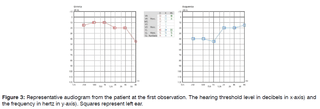
In two weeks, the patient repeated the audiogram, with a different pattern, although showing asymmetric sensorineural deafness, but suggesting a unilateral fluctuating hearing loss (Figure 3).
Figure 3: Representative audiogram from the patient at the first observation. The hearing threshold level in decibels in x-axis) and the frequency in hertz in y-axis). Squares represent left ear.
In this sense, the patient was kept under follow-up at the otolaryngology office and referred for evaluation by Neurology and Neurosurgery – with MRI and audiogram.
Bearing in mind the adequate flow of CSF and venous, there is no surgical indication for Neurosurgery, having been recommended the prosthetic adaptation with a view to optimizing the quality of life, maintainsing follow-up in the respective specialties.
Discussion
The main feature of a CMI is cervical and occipital cephalgia [7].
This is mainly seen in those who present to neurosurgical assessment. Otoneurological manifestations of CMI are also known. Fluctuating hearing loss is also described in the literature [8].
Usually, the presented clinic is credited to dysfunction in the lower cranial nerves and therefore presenting with oto-neurological symptoms such as tinnitus and vertigo. A review of 364 Chiari patients found hearing loss in 36% of patients [5]. Several case reports have been published on CMI patients with tinnitus and sensorineural hearing loss [9-12]. The literature is still very heterogeneous due to the range of the associated symptoms and respective responses to the proposed treatment. Assumptions have been suggested to clarify the development of hearing loss in CMI patients. Including stretching of the VIII nerve due to brainstem herniation, compression of the cochlear nuclei or eighth nerve by the cerebellar tonsils, ischemia of the cochlear nuclei due to effects on the posterior inferior cerebellar artery or its branches, and inner ear fluid hypertension secondary to abnormalities in cerebrospinal fluid flow [2, 9-13].
MRI is the imaging modality of choice. On sagittal imaging, the best plane for assessing the presence of MCI, the tonsils are pointed, rather than rounded and referred to as peg-like (Figure 2). According to the literature, CSF flow studies may also be useful to assess the flow surrounding the cervico-medullary junction [14]. Although described more in females, our case is also different in this sense [15]. In this case, our differential diagnosis included pseudotumor cerebri - vital as the treatment is based in a posterior fossa decompression [14]; idiopathic unilateral sudden deafness- although no response to corticosteroids was seen it could also be a possible cause for the symptomatology, Meniere syndrome – although no vestibular symptoms were shown by the patient. The presence of a CMI in an MRI and a group discussion between Neurology, Neurosurgery, and Otolaryngology postulated CMI as the most likely diagnostic and therefore follow-up was performed according to that - in three years no vestibulopathy symptoms were developed, nor an improvement in hearing. Assessment of features of intracranial hypertension is mandatory for the exclusion of pseudotumor cerebri [14]. The surgical approach of these features, by neurosurgery, is usually a suboccipital craniectomy and C1 laminectomy with Intradural exploration [16]. The patient must be informed of the operational risks and the prognosis of the disease and of the presented clinic.
Conclusion
CMI can present with numerous features, including significant hearing loss, and tinnitus - as presented in this case. CMI should be thought of as a possible etiology in anyone who presents with fluctuating hearing loss. The evaluation of the hearing status must be taken into account in the preoperative evaluation of CMI patients and can balance the surgical decision.
References
- Chiari HA. About changes in the cerebellum due to hydrocephaly of the cerebrum. DMW-German Med Weekly. 1891;17(42):1172-5.
- Rydell RE, Pulec JL. Arnold-Chiari malformation: Neuro-otologic symptoms. Arch Otolaryngol. 1971;94(1):8-12.
- Sansur CA, Heiss JD, DeVroom HL, Eskioglu E, Ennis R, Oldfield EH. Pathophysiology of headache associated with cough in patients with Chiari I malformation. J Neurosurg. 2003;98(3):453-8.
- Grahovac G, Pundy T, Tomita T. Chiari type I malformation of infants and toddlers. Childs Nerv Syst. 2018;34(6):1169-76.
- Milhorat TH, Chou MW, Trinidad EM, Kula RW, Mandell M, Wolpert C, et al. Chiari I malformation redefined: Clinical and radiographic findings for 364 symptomatic patients. Neurosurg. 1999;44(5):1005-17.
- Chandrasekhar SS, Tsai Do BS, Schwartz SR, Bontempo LJ, Faucett EA, Finestone SA, et al. Clinical practice guideline: Sudden hearing loss (update). Otolaryngol Head Neck Surg. 2019;161(1_suppl):S1-45.
- Dyste GN, Menezes AH, VanGilder JC. Symptomatic Chiari malformations: An analysis of presentation, management, and long-term outcome. J Neurosurg. 1989;71(2):159-68.
- Sperling NM, Franco Jr RA, Milhorat TH. Otologic manifestations of Chiari I malformation. Otol Neurotol. 2001;22(5):678-81.
- Ahmmed AU, Mackenzie I, Das VK, Chatterjee S, Lye RH. Audio-vestibular manifestations of Chiari malformation and outcome of surgical decompression: A case report. J Laryngol Otol. 1996;110(11):1060-4.
- Chait GE, Barber HO. Arnold-Chiari malformation--some otoneurological features. J Otolaryngol. 1979;8(1):65-70.
- Hendrix RA, Bacon CK, Sclafani AP. Chiari-I malformation associated with asymmetric sensorineural hearing loss. J Otolaryngol. 1992;21(2):102-7.
- Kumar A, Patni AH, Charbel F. The Chiari I malformation and the neurotologist. Otol Neurotol. 2002;23(5):727-35.
- Bertrand RA, Martinez SN, Françoise R. Vestibular manifestations of cerebellar ectopia. Adv Otorhinolaryngol. 1973;19:356-66.
- Strahle J, MuraSzKo KM, Kapurch J, Bapuraj JR, Garton HJ, Maher CO. Chiari malformation Type I and syrinx in children undergoing magnetic resonance imaging. J Neurosurg Pediatr. 2011;8(2):205-13.
- Ketonen LM, Hiwatashi A, Sidhu R, Westesson PL. Pediatric brain and spine: An atlas of MRI and spectroscopy. Springer Sci Business Media. 2004.
- Decq P, Le Guérinel C, Sol JC, Brugières P, Djindjian M, Nguyen JP. Chiari I malformation: a rare cause of noncommunicating hydrocephalus treated by third ventriculostomy. J Neurosurg. 2001;95(5):783-90.
1Department of Otolaryngology, Centro Hospitalar Universitário do Porto, Porto, Portugal
2Faculty of Health Sciences, Universidade da Beira Interior, Covilha, Portugal
Send correspondence to:
Andre Machado
Department of Otolaryngology, Centro Hospitalar Universitário do Porto, Porto, Portugal, E-mail: sousamachado.andre@gmail.com
Tel no: 939069577
Paper submitted on November 27, 2022; and Accepted on January 17, 2023
Citation: Machado A, Dias J, Silva A, Meireles L. Chiari Type 1 Malformation: An Unusual Cause of Tinnitus. Int Tinnitus J. 2023;27(1):06-09.


