
The International Tinnitus Journal
Official Journal of the Neurootological and Equilibriometric Society
Official Journal of the Brazil Federal District Otorhinolaryngologist Society

 44 7897 074717
ISSN: 0946-5448

Google scholar citation report
Citations : 12717
The International Tinnitus Journal received 12717 citations as per google scholar report

The International Tinnitus Journal peer review process verified at publons
Indexed In
- Excerpta Medica
- Scimago
- SCOPUS
- Publons
- EMBASE
- Google Scholar
- Euro Pub
- CAS Source Index (CASSI)
- Index Medicus
- Medline
- PubMed
- UGC
- EBSCO
Volume 30, Issue 2 / October 2026
Research Article Pages:33-38
10.5935/0946-5448.2026005
Integrated Clinical, Endoscopic, and CBCT Phenotyping of Sinonasal Disease: Diagnostic and Audiological Implications
Authors:
Alisher Mukhiddinov,Jamol Djurayev, Nargiza Normirova, Dilnoza Khusanova
Abstract
Objective: To identify clinical, endoscopic, and cone beam computed tomography features that distinguish maxillary sinus cysts from non-cystic rhinosinusitis and to describe the otologic relevance of sinonasal inflammation. Methods: This comparative observational study included 120 patients evaluated in a tertiary otorhinolaryngology service: 40 with non-cystic rhinosinusitis and 80 with maxillary sinus cysts. Symptoms, disease history, nasal endoscopy, and cone beam computed tomography findings were compared. Fisher exact tests and univariable odds ratios were recalculated from the supplied aggregate counts. Age was explored from patient-level spreadsheets after exclusion of one biologically impossible birth-year entry. Results: Cysts were more often detected incidentally (57.5% versus 20.0%; odds ratio 5.41, 95% confidence interval 2.22-13.21) and followed a prolonged asymptomatic course (61.3% versus 27.5%; odds ratio 4.17, 95% confidence interval 1.82-9.53). Diffuse mucosal thickening was less frequent in the cyst group (37.5% versus 85.0%; odds ratio 0.11, 95% confidence interval 0.04-0.28), whereas localized maxillary change was more frequent (62.5% versus 35.0%; odds ratio 3.10, 95% confidence interval 1.40-6.83). Mean lesion attenuation was lower for cysts than for inflammatory disease (+12 ± 8 versus +28 ± 12 Hounsfield units; p<0.001). Exploratory boxplots showed higher leukocyte count, erythrocyte sedimentation rate, and platelet count in the rhinosinusitis group. Conclusion: A localized, well-demarcated, low-attenuation maxillary lesion with limited endoscopic inflammation and an incidental or minimally symptomatic presentation supports a cyst diagnosis, whereas diffuse mucosal disease, discharge, and ostiomeatal obstruction favor rhinosinusitis. Prospective studies should add objective hearing, middleear, Eustachian-tube, and tinnitus outcomes.
Keywords:
Maxillary sinus cyst; rhinosinusitis; cone beam computed tomography; nasal endoscopy; Eustachian tube
dysfunction; hearing loss; tinnitus.
Introduction
Maxillary sinus cysts are common benign imaging findings. They are usually described as rounded or dome-shaped, well-circumscribed soft-tissue opacities and often arise from the sinus floor. Reported prevalence varies substantially with population and imaging modality, and cone beam computed tomography detects more lesions than panoramic radiography [1]. Long-term follow-up studies indicate that many retention cysts regress or remain stable, supporting observation when symptoms, complications, or diagnostic uncertainty are absent [2-5]. Recent surgical evidence further suggests that anatomical location may correspond to distinct histopathological phenotypes [6].
The clinically important problem is not simply recognizing a cyst, but separating a localized cystic lesion from diffuse inflammatory rhinosinusitis and from odontogenic disease. Symptoms overlap, and nasal obstruction, facial pressure, headache, discharge, and olfactory impairment are not specific [1]. Endoscopy and cross-sectional imaging therefore complement history, particularly when ostiomeatal obstruction, mucosal thickening, reduced aeration, dental pathology, or unilateral disease is present [7].
This distinction also has potential neuro-otologic relevance. Chronic sinonasal inflammation can impair Eustachian tube function and may produce aural fullness, muffled or conductive hearing loss, otalgia, and tinnitus [8-10]. Ear-related symptoms are frequent in chronic rhinosinusitis and often improve after effective sinonasal treatment, although evidence is strongest for Eustachian tube dysfunction rather than direct sensorineural injury [9]. Vascular and endothelial causes of sensorineural hearing loss and tinnitus should remain in the differential diagnosis when audiological abnormalities are present [11].
The present study compared clinical, endoscopic, and cone beam computed tomography findings in patients with maxillary sinus cysts and patients with non-cystic rhinosinusitis. A secondary objective was to define the audiological measurements required in future prospective validation without attributing unmeasured hearing or tinnitus outcomes to the current cohort [12].
Materials and Methods
Study design and participants
The clinical component was conducted at the Department of Otorhinolaryngology of the Surkhandarya Regional Multidisciplinary Medical Center during 2022-2026. The supplied cohort comprised 120 patients: 40 classified as non-cystic rhinosinusitis and 80 classified as maxillary sinus cysts [13]. Group assignment was based on clinical assessment, nasal endoscopy, and radiologic findings. The source dissertation described 70 men and 50 women overall. Patient names and medical-record identifiers were not used in this analysis or manuscript [14-20].
Patient-level spreadsheets were used only for exploratory age and mucociliary analyses [17]. Age could be calculated for all 40 non-cystic rhinosinusitis records and 79 of 80 cyst records; one cyst record contained a biologically impossible birth year and was excluded from age-based analyses. The observed age range in the spreadsheets was 13-81 years. No imputation was performed [16].
Clinical and endoscopic assessment
Clinical assessment recorded nasal obstruction, nasal discharge, headache, facial pressure or pain, olfactory dysfunction, malaise, prior treatment, recurrence, incidental detection, and prolonged asymptomatic course [21]. Rigid nasal endoscopy with 0° and 30° optics, and additional angled optics when required, assessed mucosal hyperemia, edema, pathological discharge, ostiomeatal complex obstruction, turbinate hypertrophy, and septal deviation. Local anesthesia and topical decongestion were used according to the source protocol [20].
Cone beam computed tomography assessment
Cone beam computed tomography images were reviewed in axial, sagittal, and coronal planes, with three-dimensional reconstruction when required [22]. The assessment included the distribution of mucosal disease, sinus pneumatization, ostiomeatal complex patency, localized versus diffuse change, lesion size and margins, and attenuation in Hounsfield units [23-26]. The source dataset described cysts as dome-shaped, homogeneous, and sharply marginated; mean cyst size was 12.4 ± 4.6 mm [25].
Exploratory mucociliary analysis
The saccharin transit test and nasal absorptive-function measurement were available for the cyst cohort. A saccharin particle was placed in the anterior nasal cavity and the time until perception of a sweet taste was recorded in minutes [25]. Spearman rank correlation was used to explore age versus transit time and transit time versus absorptive function. These analyses are not tests of hearing or tinnitus [27].
Audiological relevance and proposed measurement set
Hearing ability and tinnitus were not outcomes in the supplied dissertation tables or spreadsheets [24]. Accordingly, no prevalence or treatment effect for hearing loss or tinnitus was estimated. For prospective validation, the minimum audiological set should include otoscopy, pure-tone thresholds at 0.25-8 kHz, speech audiometry, tympanometry, acoustic reflexes, the Eustachian Tube Dysfunction Questionnaire 7, and documentation of aural fullness. Participants reporting tinnitus should additionally complete the Tinnitus Handicap Inventory, with tinnitus laterality, duration, and psychoacoustic characteristics recorded [28-30]. This design would distinguish middle-ear conductive effects related to Eustachian tube dysfunction from sensorineural hearing impairment [29].
Statistical analysis
Categorical variables are reported as counts and percentages. Two-sided Fisher exact tests were used for group comparisons, and univariable odds ratios with 95% confidence intervals express the odds of a cyst diagnosis when a feature was present versus absent [30]. The between-group attenuation difference was evaluated with a Welch independent-samples t test using the reported means, standard deviations, and sample sizes [31-34]. Age and laboratory distributions were compared with the Mann-Whitney U test. Spearman rank correlation assessed age versus saccharin transit time and transit time versus absorptive function in the cyst cohort. Laboratory and functional analyses were exploratory; no multiplicity-adjusted confirmatory claim was made. Statistical significance was set at p<0.05. Analyses were performed in Python 3 with SciPy [33].
Results
Statistical analysis
The median age was 34 years (interquartile range 25-38) in the non-cystic rhinosinusitis group and 33 years (interquartile range 26.5-41.5) in the cyst group. The distributions did not differ significantly (Mann-Whitney p=0.541; Figure 1).
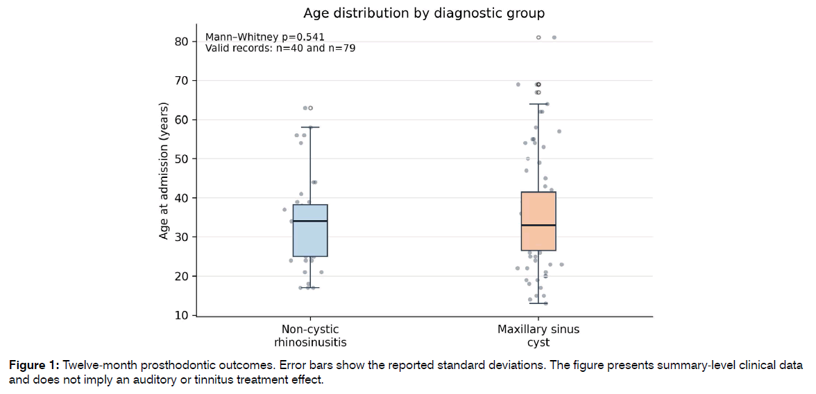
Figure 1: Twelve-month prosthodontic outcomes. Error bars show the reported standard deviations. The figure presents summary-level clinical data and does not imply an auditory or tinnitus treatment effect.
Clinical presentation
Inflammatory sinonasal symptoms were consistently more frequent in non-cystic rhinosinusitis. Nasal discharge, nasal obstruction, olfactory dysfunction, headache, and malaise were negatively associated with a cyst diagnosis. Facial pressure or pain alone did not distinguish the groups. Conversely, incidental detection and a prolonged asymptomatic course were strongly associated with cysts (Table 1).
| Feature | Rhinosinusitis n=40 | Cyst n=80 | OR for cyst diagnosis 95% CI |
|---|---|---|---|
| Nasal obstruction | 35 (87.5) | 42 (52.5) | 0.16 (0.06-0.44); p<0.001 |
| Nasal discharge | 32 (80.0) | 27 (33.8) | 0.13 (0.05-0.31); p<0.001 |
| Headache | 27 (67.5) | 32 (40.0) | 0.32 (0.14-0.71); p=0.006 |
| Facial pressure or pain | 25 (62.5) | 38 (47.5) | 0.54 (0.25-1.18); p=0.174 |
| Olfactory dysfunction | 24 (60.0) | 17 (21.2) | 0.18 (0.08-0.41); p<0.001 |
| Malaise | 19 (47.5) | 20 (25.0) | 0.37 (0.17-0.82); p=0.022 |
| Incidental detection | 8 (20.0) | 46 (57.5) | 5.41 (2.22-13.21); p<0.001 |
| Prolonged asymptomatic course | 11 (27.5) | 49 (61.3) | 4.17 (1.82-9.53); p<0.001 |
Table 1: Clinical features by diagnostic group
Exploratory laboratory profiles
Leukocyte counts were higher in non-cystic rhinosinusitis than in the cyst group (median 11.9 versus 7.04 ×10⁹/L; p<0.001). Erythrocyte sedimentation rate was also higher (median 13 versus 8 mm/h; p<0.001), as were platelet counts (median 355 versus 265.5 ×10⁹/L; p<0.001; Figure 2). These post hoc findings describe the supplied cohort and require independent confirmation.
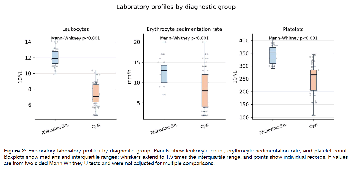
Figure 2: Exploratory laboratory profiles by diagnostic group. Panels show leukocyte count, erythrocyte sedimentation rate, and platelet count. Boxplots show medians and interquartile ranges; whiskers extend to 1.5 times the interquartile range, and points show individual records. P values are from two-sided Mann-Whitney U tests and were not adjusted for multiple comparisons.
Endoscopic findings
Mucosal hyperemia, edema, pathological discharge, ostiomeatal complex obstruction, and turbinate hypertrophy were less frequent in the cyst group. Septal deviation was common in both groups and was not statistically discriminative (Table 2).
| Feature | Rhinosinusitis n=40 | Cyst n=80 | OR for cyst diagnosis 95% CI |
|---|---|---|---|
| Mucosal hyperemia | 33 (82.5) | 33 (41.2) | 0.15 (0.06-0.38); p<0.001 |
| Mucosal edema | 31 (77.5) | 31 (38.8) | 0.18 (0.08-0.44); p<0.001 |
| Pathological discharge | 30 (75.0) | 26 (32.5) | 0.16 (0.07-0.38); p<0.001 |
| Ostiomeatal complex obstruction | 29 (72.5) | 30 (37.5) | 0.23 (0.10-0.52); p<0.001 |
| Turbinate hypertrophy | 24 (60.0) | 29 (36.2) | 0.38 (0.17-0.83); p=0.019 |
| Septal deviation | 24 (60.0) | 34 (42.5) | 0.49 (0.23-1.07); p=0.083 |
Table 2: Endoscopic features by diagnostic group
Computed tomographic findings
Diffuse mucosal thickening, reduced pneumatization, ostiomeatal complex obstruction, and turbinate hypertrophy favored non-cystic rhinosinusitis, whereas localized maxillary change favored a cyst diagnosis. Septal deviation again did not distinguish the groups. Mean attenuation was +28 ± 12 Hounsfield units in rhinosinusitis and +12 ± 8 Hounsfield units in cysts; the difference was significant (Welch t=7.63, p<0.001) (Table 3).
| Feature | Rhinosinusitis n=40 | Cyst n=80 | Association or comparison |
|---|---|---|---|
| Diffuse mucosal thickening | 34 (85.0) | 30 (37.5) | 0.11 (0.04-0.28); p<0.001 |
| Reduced sinus pneumatization | 31 (77.5) | 28 (35.0) | 0.16 (0.07-0.37); p<0.001 |
| Ostiomeatal complex obstruction | 29 (72.5) | 33 (41.2) | 0.27 (0.12-0.61); p=0.002 |
| Localized maxillary change | 14 (35.0) | 50 (62.5) | 3.10 (1.40-6.83); p=0.006 |
| Septal deviation | 25 (62.5) | 36 (45.0) | 0.49 (0.23-1.07); p=0.083 |
| Turbinate hypertrophy | 23 (57.5) | 27 (33.8) | 0.38 (0.17-0.82); p=0.018 |
| Mean attenuation HU | +28 ± 12 | +12 ± 8 | Welch t=7.63; p<0.001 |
Table 3: Cone beam computed tomography features by diagnostic group
Exploratory mucociliary finding
Among 79 cyst records with valid age and saccharin transit data, age was not associated with mucociliary clearance time (Spearman ρ=-0.09, p=0.443; Figure 3). Mucociliary clearance time was likewise not associated with absorptive function (Spearman ρ=0.05, p=0.639; Figure 4). These null exploratory findings do not establish normal nasal physiology and should not be extrapolated to the rhinosinusitis group, for which equivalent patient-level functional data were not available in the supplied spreadsheet (Figure 3,4).
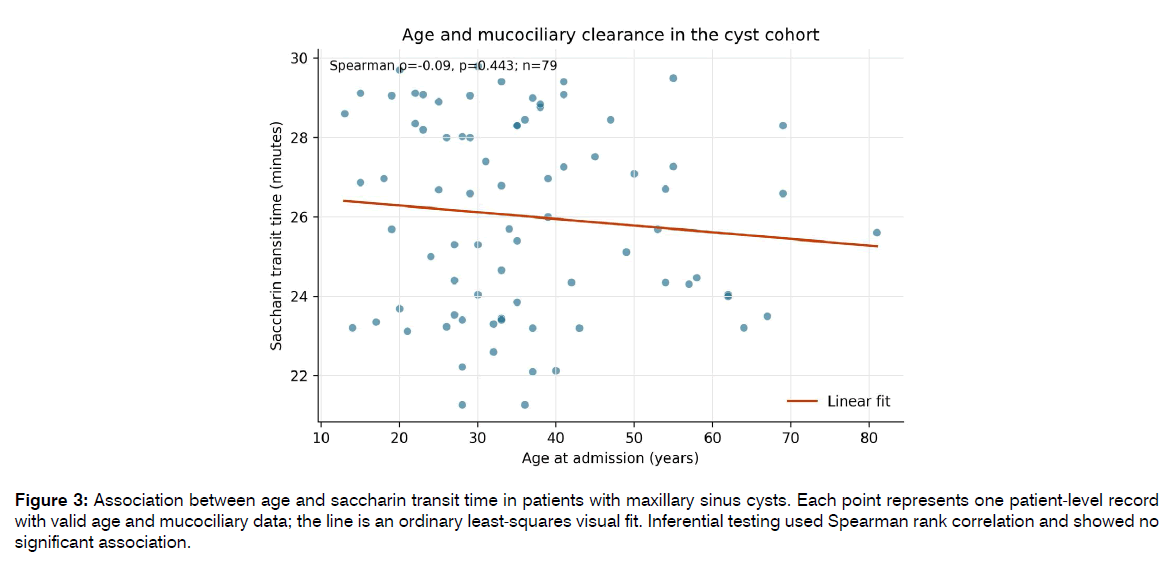
Figure 3: Association between age and saccharin transit time in patients with maxillary sinus cysts. Each point represents one patient-level record with valid age and mucociliary data; the line is an ordinary least-squares visual fit. Inferential testing used Spearman rank correlation and showed no significant association.
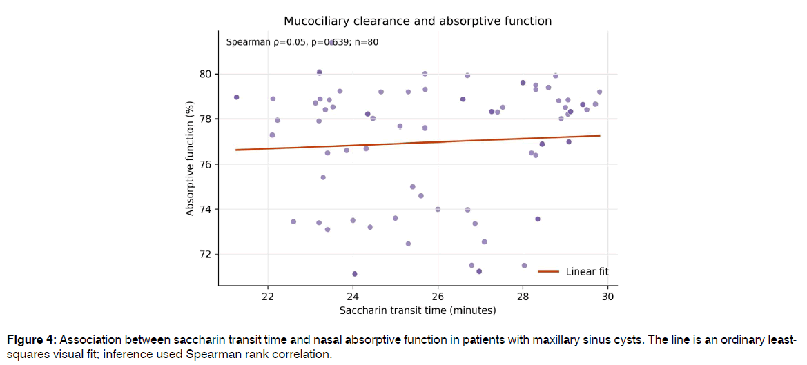
Figure 4: Association between saccharin transit time and nasal absorptive function in patients with maxillary sinus cysts. The line is an ordinary leastsquares visual fit; inference used Spearman rank correlation.
Discussion
The study identifies a coherent pattern that separates maxillary sinus cysts from non-cystic rhinosinusitis [35]. Cysts were more likely to be incidental, minimally symptomatic, localized, sharply marginated, and lower in attenuation. Non-cystic rhinosinusitis showed diffuse inflammatory change across history, endoscopy, and imaging [36]. The strongest positive clinical associations with a cyst diagnosis were incidental detection and prolonged asymptomatic course; the strongest negative associations were diffuse mucosal thickening, pathological discharge, hyperemia, and edema.
These findings agree with earlier radiographic and cone beam computed tomography studies in which mucous retention cysts were frequently asymptomatic and located along the maxillary sinus floor, while their observed prevalence varied with imaging technique and population [37-40]. They also support the principle that imaging should be interpreted in clinical context. A dome-shaped opacity is not automatically the cause of facial pain or headache, and diffuse inflammatory changes or a dental source may require a different diagnostic pathway [38].
Recent evidence from 140 surgically treated cases further indicates that imaging-defined cyst location may correspond to distinct epithelial, inflammatory, and stromal histopathological phenotypes [39,40]. This supports location-aware assessment and targeted tissue sampling while retaining histopathology as the definitive method when excision is undertaken [35].
The attenuation difference is clinically attractive because it is objective; however, Hounsfield values on cone beam computed tomography are not as standardized as values on conventional multidetector computed tomography [41-45]. Scanner settings, reconstruction, field of view, artifacts, and calibration can shift gray values. The reported +12 versus +28 Hounsfield-unit contrast should therefore be treated as an internal cohort finding rather than a universal diagnostic threshold [42]. External validation should use prespecified acquisition parameters, blinded readers, and conventional CT calibration or phantom correction [46].
The higher leukocyte count, erythrocyte sedimentation rate, and platelet count in the rhinosinusitis comparator are biologically compatible with a more inflammatory phenotype [47,48]. However, these variables were selected after inspection of the available spreadsheet, treatment status and comorbidities were not modeled, and multiple laboratory comparisons were not corrected [43]. The boxplots should therefore be interpreted as descriptive, hypothesis-generating visualizations rather than diagnostic cutoffs.
Audiological relevance
The present cohort cannot answer whether maxillary sinus cysts cause hearing impairment or tinnitus [49,50]. Nevertheless, the comparison is relevant to an otology-focused readership because the inflammatory phenotype showed more ostiomeatal obstruction, edema, and discharge, which may coexist with nasopharyngeal inflammation and Eustachian tube dysfunction [30]. Meta-analytic evidence suggests that approximately half of selected patients with chronic rhinosinusitis may meet a symptom threshold consistent with Eustachian tube dysfunction, and several studies have reported improvement in ear fullness and related otologic symptoms after endoscopic sinus surgery [51]. Eustachian tube dysfunction can produce negative middle-ear pressure, conductive hearing loss, aural fullness, and tinnitus; it should not be equated with sensorineural hearing loss without audiometry and tympanometry [17].
A 2026 population-based analysis also reported an association between prior chronic rhinosinusitis or allergic rhinitis and tinnitus, but observational association does not establish that a maxillary sinus cyst causes tinnitus. A defensible follow-up study should compare cyst and rhinosinusitis groups using pure-tone average, air-bone gaps, tympanometric type, ETDQ-7, and THI at baseline and after treatment [52]. This would align the rhinologic question with the International Tinnitus Journal while preserving causal and measurement discipline.
The differential diagnosis should also consider non-rhinologic causes of sensorineural hearing loss and tinnitus. Shodmonkulova and colleagues have demonstrated the clinical relevance of vascular and endothelial mechanisms in auditory dysfunction and emphasized combined audiometric, tinnitus, and vascular assessment in appropriately selected patients [33].
Strengths and limitations
Strengths include direct comparison of two clinically overlapping sinonasal conditions, concordant assessment across symptoms, endoscopy, and imaging, and effect estimates that quantify rather than merely describe group differences. The study also avoids claiming audiological outcomes that were not measured [53].
The main limitations are the single-center observational design, unequal group sizes, absence of a prespecified diagnostic model or external validation cohort, and lack of patient-level data for several aggregate variables. Sex-specific and group-specific age summaries were not consistently available across source tables. One biologically impossible birth-year value was excluded from age analyses [54]. The rhinosinusitis spreadsheet included heterogeneous diagnostic labels, so the comparator is described conservatively as non-cystic rhinosinusitis. Individual computed tomographic attenuation values were unavailable; therefore, no attenuation boxplot or patient-level receiver operating characteristic analysis was possible. Laboratory visualizations were post hoc and potentially confounded, while functional scatter plots were restricted to the cyst cohort. Finally, no otoscopy, pure-tone audiometry, tympanometry, ETDQ-7, THI, or tinnitus phenotype was collected, preventing conclusions about hearing or tinnitus [53,54].
Conclusion
Maxillary sinus cysts are distinguished from non-cystic rhinosinusitis by an incidental or prolonged asymptomatic course, limited endoscopic inflammation, localized imaging change, and lower mean lesion attenuation. Diffuse mucosal thickening, reduced pneumatization, pathological discharge, and ostiomeatal obstruction favor inflammatory rhinosinusitis. These findings support integrated clinical, endoscopic, and imaging interpretation rather than reliance on a single symptom or opacity. Prospective validation should incorporate objective hearing, middle-ear, Eustachian-tube, and tinnitus measures before any audiological claim is made.
References
- Fokkens WJ, Lund VJ, Hopkins C, et al. European Position Paper on Rhinosinusitis and Nasal Polyps 2020. Rhinology. 2020;58(Suppl S29):1-464.
- Orlandi RR, Kingdom TT, Smith TL, et al. International consensus statement on allergy and rhinology: rhinosinusitis 2021. Int Forum Allergy Rhinol. 2021;11(3):213-739.
- Rosenfeld RM, Piccirillo JF, Chandrasekhar SS, et al. Clinical practice guideline update: adult sinusitis. Otolaryngol Head Neck Surg. 2015;152(2 Suppl):S1-S39.
- Giotakis EI, Weber RK. Cysts of the maxillary sinus: a literature review. Int Forum Allergy Rhinol. 2013;3:766-771.
- Wang JH, Jang YJ, Lee BJ. Natural course of retention cysts of the maxillary sinus: long-term follow-up results. Laryngoscope. 2007;117:341-344.
- Vieira EMM, de Morais S, Musis CR, et al. Frequency of maxillary sinus mucous retention cysts in a central Brazilian population. J Dent Shiraz Univ Med Sci. 2015;16:169-174.
- Ren L, Chen C, Li N, et al. Prevalence of and factors associated with maxillary sinus cyst in a Chinese population. J Oral Sci. 2022;64:22-27.
- Ruprecht A, Batniji S, el-Neweihi E. Mucous retention cyst of the maxillary sinus. Oral Surg Oral Med Oral Pathol. 1986;62:728-731.
- Mathew AL, Pai KM, Sholapurkar AA. Maxillary sinus findings in the elderly: a panoramic radiographic study. J Contemp Dent Pract. 2009;10:E041-E048.
- Bhattacharyya N. Do maxillary sinus retention cysts reflect obstructive sinus phenomena? Arch Otolaryngol Head Neck Surg. 2000;126:1369-1371.
- Mardinger O, Manor I, Mijiritsky E, et al. Maxillary sinus augmentation in the presence of antral pseudocyst: a clinical approach. Oral Surg Oral Med Oral Pathol Oral Radiol Endod. 2007;103:180-184.
- Rodrigues CD, Freire GF, Silva LB, et al. Prevalence and risk factors of mucous retention cysts in a Brazilian population. Dentomaxillofac Radiol. 2009;38:480-483.
- Ramesh A, Pabla T. Mucous retention cyst of maxillary sinuses. J Mass Dent Soc. 2008;57:14-15.
- Veltrini V, Ferreira Junior O, Oliveira DT. Mucosal cysts of the maxillary sinus: a literature review. Med Oral. 2001;6:180-188.
- Khasanov U, Khasanova L, Djuraev J, et al. Anatomical location predicts histopathological phenotype in maxillary sinus cysts: a secondary analysis of 140 surgical cases. Int Tinnitus J. 2026;30(2):1-7.
- Martinez-Gonzalez JM, Barona-Dorado C, Arias-Irimia O, et al. Panoramic and tomographic implant studies: role in the diagnosis of sinus disorders. Med Oral Patol Oral Cir Bucal. 2010;15:e611-e615.
- Bosio JA, Tanaka O, Rovigatti E, et al. The incidence of maxillary sinus retention cysts in orthodontic patients. World J Orthod. 2009;10:e7-e8.
- Donizeth-Rodrigues C, Fonseca-Da Silveira M, Goncalves-De Alencar AH, et al. Three-dimensional images contribute to the diagnosis of mucous retention cyst in maxillary sinus. Med Oral Patol Oral Cir Bucal. 2013;18:e151-e157.
- MacDonald-Jankowski DS. Mucosal antral cysts in a Chinese population. Dentomaxillofac Radiol. 1993;22:208-210.
- MacDonald-Jankowski DS. Mucosal antral cysts observed within a London inner-city population. Clin Radiol. 1994;49:195-198.
- Rege IC, Sousa TO, Leles CR, et al. Occurrence of maxillary sinus abnormalities detected by cone beam CT in asymptomatic patients. BMC Oral Health. 2012;12:30.
- Bell GW, Joshi BB, Macleod RI. Maxillary sinus disease: diagnosis and treatment. Br Dent J. 2011;210:113-118.
- Vallo J, Suominen-Taipale L, Huumonen S, et al. Prevalence of mucosal abnormalities of the maxillary sinus and their relationship to dental disease in panoramic radiography. Oral Surg Oral Med Oral Pathol Oral Radiol Endod. 2010;109:e80-e87.
- Maestre-Ferrin L, Galan-Gil S, Carrillo-Garcia C, et al. Radiographic findings in the maxillary sinus: comparison of panoramic radiography with computed tomography. Int J Oral Maxillofac Implants. 2011;26:341-346.
- Carter LC, Calamel A, Haller A, et al. Seasonal variation in maxillary antral pseudocysts in a general clinic population. Dentomaxillofac Radiol. 1998;27:22-24.
- Casamassimo PS, Lilly GE. Mucosal cysts of the maxillary sinus: a clinical and radiographic study. Oral Surg Oral Med Oral Pathol. 1980;50:282-286.
- Niknami M, Mirmohammadi M, Pezeshki A. Evaluation of the prevalence of mucous retention pseudocyst and its correlation with associated risk factors using panoramic radiography and cone-beam computed tomography. J Dent Tehran. 2018;15:123-129.
- Craig JR. Odontogenic sinusitis: a state-of-the-art review. World J Otorhinolaryngol Head Neck Surg. 2022;8:8-15.
- Psillas G, Papaioannou D, Petsali S, et al. Odontogenic maxillary sinusitis: a comprehensive review. J Dent Sci. 2021;16:474-481.
- Simuntis R, Kubilius R, Vaitkus S. Odontogenic maxillary sinusitis: a review. Stomatologija. 2014;16:39-43.
- Craig JR, Tataryn RW, Aghaloo TL, et al. Management of odontogenic sinusitis: multidisciplinary consensus statement. Int Forum Allergy Rhinol. 2020;10:901-912.
- Lin J, Wang C, Wang X, et al. Expert consensus on odontogenic maxillary sinusitis multidisciplinary treatment. Int J Oral Sci. 2024;16:11.
- Mehra P, Murad H. Maxillary sinus disease of odontogenic origin. Otolaryngol Clin North Am. 2004;37:347-364.
- Brook I. Sinusitis of odontogenic origin. Otolaryngol Head Neck Surg. 2006;135:349-355.
- Zirk M, Dreiseidler T, Pohl M, et al. Odontogenic sinusitis maxillaris: a retrospective study of 121 cases with surgical intervention. J Craniomaxillofac Surg. 2017;45:520-525.
- Shodmonkulova G, Khaydarova G, Yulbarisov A, et al. Carotid artery stenosis and auditory dysfunction: a systematic review of mechanisms and interventions. Acta Otorrinolaringol Esp (Engl Ed). 2026;77(4):512331. doi:10.1016/j.otoeng.2026.512331.
- Lohiya SS, Patel SV, Pawde AM, et al. Comparative study of diagnostic nasal endoscopy and CT paranasal sinuses in diagnosing chronic rhinosinusitis. Indian J Otolaryngol Head Neck Surg. 2016;68:224-229.
- Stankiewicz JA, Chow JM. Nasal endoscopy and the definition and diagnosis of chronic rhinosinusitis. Otolaryngol Head Neck Surg. 2002;126:623-627.
- Wuister AMH, Goto NA, Oostveen EJ, et al. Nasal endoscopy is recommended for diagnosing adults with chronic rhinosinusitis. Otolaryngol Head Neck Surg. 2014;150:359-364.
- Teo NW, Mace JC, Smith TL, et al. Impact of endoscopic sinus surgery on otologic symptoms associated with chronic rhinosinusitis. World J Otorhinolaryngol Head Neck Surg. 2017;3:24-31.
- Maniakas A, Desrosiers M, Asmar MH, et al. Eustachian tube symptoms are frequent in chronic rhinosinusitis and respond well to endoscopic sinus surgery. Rhinology. 2018;56:118-121.
- Chang MT, Hosseini DK, Song SH, et al. The effect of endoscopic sinus surgery on Eustachian tube dysfunction symptoms. Otolaryngol Head Neck Surg. 2020;163:603-610.
- Chen T, Shih MC, Edwards TS, et al. Eustachian tube dysfunction in chronic rhinosinusitis with comparison to primary Eustachian tube dysfunction: a systematic review and meta-analysis. Int Forum Allergy Rhinol. 2022;12:942-951.
- Schilder AGM, Bhutta MF, Butler CC, et al. Eustachian tube dysfunction: consensus statement on definition, types, clinical presentation and diagnosis. Clin Otolaryngol. 2015;40:407-411.
- Higgins TS, Cappello ZJ, Wu AW, et al. Predictors of Eustachian tube dysfunction improvement and normalization after endoscopic sinus surgery. Laryngoscope. 2020;130:E721-E726.
- Leo G, Piacentini E, Incorvaia C, et al. Sinusitis and Eustachian tube dysfunction in children. Pediatr Allergy Immunol. 2007;18(Suppl 18):35-39.
- Stoikes NF, Dutton JM. The effect of endoscopic sinus surgery on symptoms of Eustachian tube dysfunction. Am J Rhinol. 2005;19:199-202.
- Bhattacharyya N. Symptom outcomes after endoscopic sinus surgery for chronic rhinosinusitis. Arch Otolaryngol Head Neck Surg. 2004;130:329-333.
- DeConde AS, Bodner TE, Mace JC, et al. Response shift in quality of life after endoscopic sinus surgery for chronic rhinosinusitis. JAMA Otolaryngol Head Neck Surg. 2014;140:712-719.
- Calvo-Henriquez C, Di Stadio A, Chiesa-Estomba C, et al. Pathophysiological link between chronic rhinosinusitis and ear disease. Curr Allergy Asthma Rep. 2023;23:409-417.
- Yang TH, Chung SD, Lin HC, et al. Associations of chronic rhinosinusitis and allergic rhinitis with tinnitus. Sci Rep. 2026;16:8169.
- Shodmonkulova G, Khaydarova G, Khabilov N, et al. Hearing loss in carotid artery stenosis: influence of age and gender. Int Tinnitus J. 2025;29(2):71-75. doi:10.5935/0946-5448.2025011.
- Arias-Irimia O, Barona-Dorado C, Santos-Marino JA, et al. Meta-analysis of the etiology of odontogenic maxillary sinusitis. Med Oral Patol Oral Cir Bucal. 2010;15:e70-e73.
- Shodmonkulova G, Khaydarova G, Yulbarisov A, et al. Vascular endothelial dysfunction as a mechanism of hearing loss in carotid artery stenosis: a scientific literature review. Egypt J Otolaryngol. 2026;42:106. doi:10.1186/s43163-026-01092-6.
1Department of Otorhinolaryngology, Tashkent State Medical University, Tashkent, Uzbekistan.
2Termez Branch of Tashkent State Medical University,Tashkent, Uzbekistan.
Send correspondence to:
Alisher Mukhiddinov
Department of Otorhinolaryngology, Tashkent State Medical University, Tashkent, Uzbekistan. Termez Branch of Tashkent State Medical University,Tashkent, Uzbekistan, E-mail: entsurgeon@bk.ru
Tel:+ 998 915079162
Paper submitted on Sept 23, 2026; and Accepted on Sept 28, 2026
Citation: Alisher M. Integrated Clinical, Endoscopic, and CBCT Phenotyping of Sinonasal Disease: Diagnostic and Audiological Implications. Int Tinnitus J. 2026;30(2):33-40