The International Tinnitus Journal
Official Journal of the Neurootological and Equilibriometric Society
Official Journal of the Brazil Federal District Otorhinolaryngologist Society

 44 7897 074717
ISSN: 0946-5448

Google scholar citation report
Citations : 12717
The International Tinnitus Journal received 12717 citations as per google scholar report

The International Tinnitus Journal peer review process verified at publons
Indexed In
- Excerpta Medica
- Scimago
- SCOPUS
- Publons
- EMBASE
- Google Scholar
- Euro Pub
- CAS Source Index (CASSI)
- Index Medicus
- Medline
- PubMed
- UGC
- EBSCO
Volume 30, Issue 2 / September 2026
Research Article Pages:27-32
10.5935/0946-5448.2026004
PROSTHETIC OCCLUSAL STABILITY, MASTICATORY FUNCTION, AND AUDITORY PHENOTYPING IN REMOVABLE DENTURE USERS
Authors:
Gulbonu Yahyoyeva, Bekhzod Khabilov, Firuza Mirsalikhova, Gulruh Shodmonkulova, Muyassar Mirkhoshimova, Dilnoza Sharipova, Nargiza Karimova, Shoira Khudayberganova, Shaxriyor Normirzayev, Mohinur Yahyoyeva
Abstract
Background: Somatosensory tinnitus may coexist with temporomandibular disorders and altered jaw function, while removable prostheses can change occlusal contacts and masticatory loading. Direct evidence linking artificial-tooth wear or denture-related occlusal instability to tinnitus remains limited. Objective: To examine longitudinal prosthodontic outcomes and evaluate an age-stratified audiometry analysis framework relevant to somatosensory tinnitus research. Methods: Summary-level data from a 12-month prospective prosthodontic cohort of 90 removable denture users were evaluated together with a separate simulated audiometry dataset of 90 age-stratified records used for methodological validation. Clinical outcomes included two-colour chewing-gum mixing efficiency at 15 and 30 seconds, threedimensional occlusal surface change, Vickers microhardness, and compressive strength. The audiometry dataset contained bilateral thresholds at 1000, 2000, 4000, and 8000 Hz. Boxplots and age-stratified inferential tests were used to evaluate the proposed auditory analysis pipeline; no patient-level linkage between the two datasets was assumed. Results: In the prosthodontic cohort summary, 15-second masticatory efficiency declined from 69.77 +/- 5.34% at baseline to 66.22 +/- 5.04% at 12 months, while the 30-second value declined from 87.73 +/- 6.41% to 85.01 +/- 5.17%. Mean occlusal surface change increased from 0% to 1.39 +/- 0.32%. Wear was inversely associated with masticatory efficiency (r=-0.72 for 15 seconds and r=-0.68 for 30 seconds). In the audiometry pipeline analysis, thresholds increased with age and frequency. Tinnitus-specific associations were not estimated because tinnitus endpoints were not included in that dataset. Conclusion: Longitudinal prosthodontic phenotyping can quantify clinically relevant changes in occlusal stability and mastication, but these data alone cannot establish a somatosensory tinnitus mechanism. A valid clinical study must link patient-level denture, DC/TMD, audiometric, tympanometric, and THI/TFI measurements at common time points and control strongly for age and hearing loss.
Keywords:
Removable denture; occlusal wear; masticatory performance; somatosensory tinnitus; temporomandibular
disorders; pure-tone audiometry.
Keywords
removable denture; occlusal wear; masticatory performance; somatosensory tinnitus; temporomandibular disorders; pure-tone audiometry
Introduction
Tinnitus is the perception of sound without a corresponding external acoustic source and is heterogeneous in mechanism, clinical burden, and response to treatment. Population estimates vary because case definitions and time frames differ, but contemporary syntheses confirm that tinnitus is common and that a smaller clinically important subgroup experiences persistent or disabling symptoms [1, 2]. The auditory system remains central to most tinnitus models; however, somatosensory input from the cervical and trigeminal systems can alter activity within auditory pathways and modulate tinnitus loudness or pitch in selected patients [1].
Temporomandibular disorders (TMDs), masticatory muscle pain, oral parafunctions, and tinnitus frequently coexist [3]. An updated 2026 meta-analysis reported a pooled tinnitus prevalence of 31.8% among patients with TMDs and an odds ratio of 3.924 for tinnitus in individuals with versus without TMD, while emphasizing extreme heterogeneity, observational designs, self-reported outcomes, and the inability to infer causality [4]. Earlier systematic reviews reached the same cautious conclusion: the association is clinically meaningful enough to investigate but is vulnerable to referral bias, diagnostic inconsistency, age-related confounding, and reverse causation [5, 6].
The potential relevance of removable prosthodontics is plausible but under-tested. Artificial teeth undergo material-dependent wear; changes in cusp-fossa anatomy and occlusal contacts may alter masticatory muscle recruitment, mandibular position, prosthesis stability, and symptom-provoking jaw movements. Clinical studies have linked poor prosthesis occlusion or vertical dimension to worse masticatory performance [7], and optical three-dimensional methods can quantify denture-tooth wear over time [8]. These observations do not prove a tinnitus mechanism, but they identify measurable prosthodontic exposures that could be incorporated into a rigorous somatosensory tinnitus study.
The analysis included longitudinal summary outcomes from a cohort of 90 removable denture users followed for 12 months, with repeated masticatory-performance and three-dimensional occlusal-surface measurements. A separate simulated audiometry dataset of 90 age-stratified records was used solely to evaluate the statistical and visualization workflow. Because the datasets were not linked at participant level, the auditory analysis is presented as methodological validation and not as evidence of an association between denture-related outcomes, hearing thresholds, or tinnitus [9].
Methods
Study design and data structure
This methodological integration study used aggregate outcomes from a prospective 12-month clinical cohort of 90 adults with partial secondary edentulism treated with removable acrylic plate dentures. A separate simulated audiometry dataset contained 90 records divided equally among three age strata and was used only to evaluate the analysis workflow. The datasets had no common participant identifier and were not statistically linked [10].
Clinical prosthodontic cohort
The prosthodontic cohort comprised 90 patients aged 18-74 years, with 30 participants in each of the 18-44, 45-59, and 60-74 year groups. All underwent removable plate-denture rehabilitation and were evaluated at baseline, 6 months, and 12 months. Only aggregate cohort outcomes were available for the present analysis; participant-level demographic data, recruitment dates, and detailed eligibility criteria were not available for independent reanalysis [9, 10].
Masticatory performance
Masticatory performance was assessed using a two-colour chewing-gum mixing test at 15 and 30 seconds. After chewing, samples were photographed and processed by an automated hue-saturation-value colour-segmentation method that quantified the proportion and uniformity of mixed colour zones. The method is described as automated colour segmentation rather than a machine-learning algorithm. Two-colour mixing tests are established tools for quantifying masticatory function, including in complete-denture wearers, when image acquisition and analysis are standardized [11].
Occlusal surface change and material properties
Artificial-tooth occlusal surfaces were scanned at baseline, 6 months, and 12 months using a Medit T710 laboratory scanner. Digital models were compared in exocad, and surface change was expressed as a percentage. The laboratory component measured Vickers microhardness under an HV 0.5 protocol and compressive strength. These measurements characterize material behavior and occlusal stability; they do not by themselves diagnose TMD or somatosensory tinnitus [12].
Audiometry analysis framework
The audiometry pipeline dataset included right- and left-ear air-conduction thresholds at 1000, 2000, 4000, and 8000 Hz. Binaural frequency-specific values were calculated as the arithmetic mean of the two ears. Right-ear and left-ear four-frequency averages and the lower (better-ear) average were also available. The term 'PTA4' was not used because 500 Hz was unavailable; the metric was therefore labelled as an average from 1-8 kHz. Tinnitus presence and THI/TFI scores were not included in the pipeline dataset.
Statistical analysis
Clinical prosthodontic outcomes were summarized using means, standard deviations, and reported correlations. Because individual clinical observations were unavailable for reanalysis, repeated-measures models, distribution checks, attrition analyses, and adjusted associations could not be independently reproduced. For the simulated audiometry pipeline dataset, frequency-specific distributions were summarized by mean, standard deviation, median, interquartile range, minimum, and maximum. One-way ANOVA and Kruskal-Wallis tests compared the three age strata; these tests evaluated the behavior of the analytical pipeline and are not interpreted as clinical evidence. Boxplots show medians, interquartile ranges, whiskers, and outliers. Analyses were performed using pandas, SciPy, NumPy, and Matplotlib [13].
Results
Clinical prosthodontic outcomes
Masticatory efficiency decreased modestly across 12 months. At 15 seconds, the mean declined from 69.77 +/- 5.34% at baseline to 67.89 +/- 5.30% at 6 months and 66.22 +/- 5.04% at 12 months. At 30 seconds, the corresponding means were 87.73 +/- 6.41%, 86.35 +/- 5.66%, and 85.01 +/- 5.17%. The longer chewing interval consistently produced higher mixing values, but both series showed the same direction of change.
Mean occlusal surface change increased from 0% at baseline to 0.68 +/- 0.20% at 6 months and 1.39 +/- 0.32% at 12 months. The dissertation summary reported inverse correlations between surface change and masticatory efficiency (r=-0.72 for the 15-second test and r=-0.68 for the 30-second test; both p<0.001). These estimates support internal coherence between progressive wear and reduced mixing performance, but confidence intervals and adjusted models could not be recalculated without participant-level data Table 1, Figure 1.
| Outcome | Baseline | 6 months | 12 months |
|---|---|---|---|
| Masticatory efficiency, 15 seconds (%) | 69.77 +/- 5.34 | 67.89 +/- 5.30 | 66.22 +/- 5.04 |
| Masticatory efficiency, 30 seconds (%) | 87.73 +/- 6.41 | 86.35 +/- 5.66 | 85.01 +/- 5.17 |
| Occlusal surface change (%) | 0 | 0.68 +/- 0.20 | 1.39 +/- 0.32 |
Table 1: Twelve-month prosthodontic outcomes
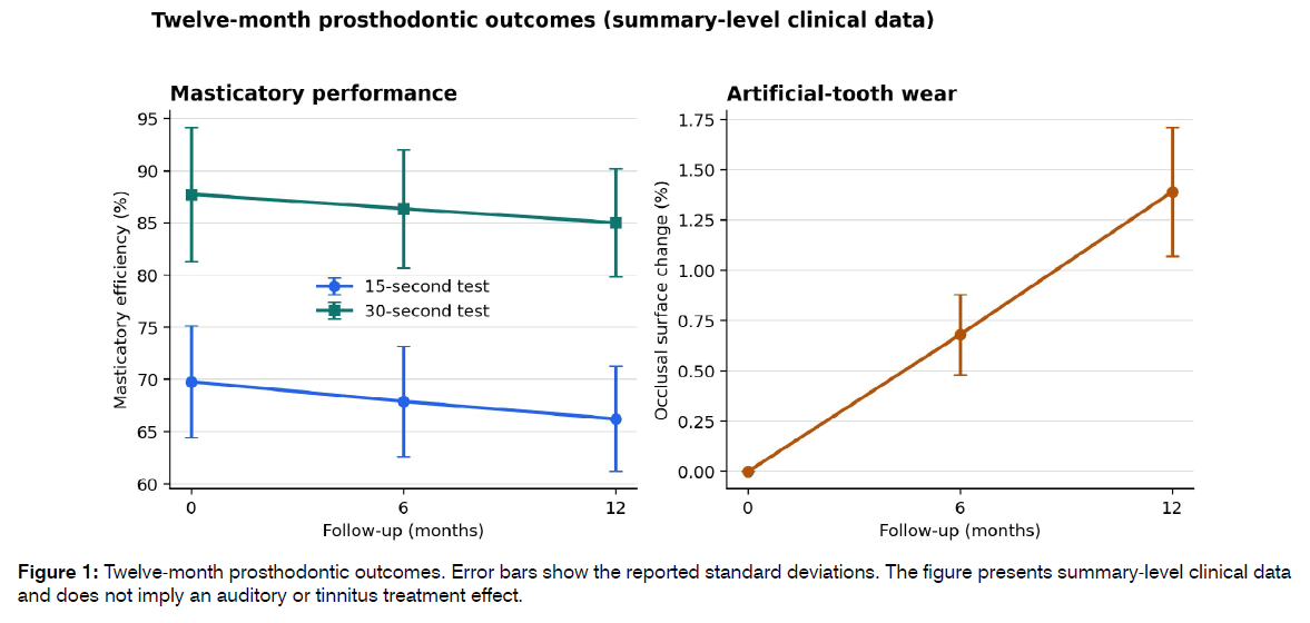
Figure 1: Twelve-month prosthodontic outcomes. Error bars show the reported standard deviations. The figure presents summary-level clinical data and does not imply an auditory or tinnitus treatment effect.
Material-specific findings
Microhardness varied widely across the evaluated materials. Eray had the highest reported value (176.1 HV) and the lowest 12-month surface change (1.05 +/- 0.25%). Yamahachi showed the highest compressive strength (135 +/- 4.5 MPa) but greater 12-month surface change (1.55 +/- 0.30%). The source summary reported correlations of r=0.74 between microhardness and preservation of the occlusal surface and r=0.61 between compressive strength and surface preservation. These results reinforce the need to evaluate hardness, strength, wear, occlusal behavior, and function jointly rather than treating a single material property as a surrogate for clinical benefit Table 2.
| Material | Vickers microhardness (HV) | Compressive strength (MPa) | 12-month surface change (%) |
|---|---|---|---|
| Tiziano | 40.1 | 130 +/- 4.2 | 1.42 +/- 0.29 |
| Yamahachi | 27.0 | 135 +/- 4.5 | 1.55 +/- 0.30 |
| Super S | 23.9 | 132 +/- 4.3 | 1.60 +/- 0.31 |
| Eray | 176.1 | 120 +/- 3.8 | 1.05 +/- 0.25 |
| Kali | 67.9 | 115 +/- 4.0 | 1.45 +/- 0.28 |
| Press Ceramic | 56.3 | 128 +/- 4.1 | Not reported |
| Spofad | 22.6 | Not reported | 1.48 +/- 0.27 |
Table 2: Artificial-tooth material properties and 12-month surface change
Audiometry analysis pipeline
The simulated pipeline dataset contained 90 records, with 30 records per age stratum; 56 were labelled female and 34 male. The hearing-category field classified 40 records as normal, 42 as mild, and 8 as moderate. These methodological categories do not estimate hearing-loss prevalence among removable denture users.
The pipeline dataset showed an age-frequency gradient. Mean binaural thresholds in the 18-44, 45-59, and 60-74 year groups were 13.1, 18.5, and 26.2 dB HL at 1000 Hz; 14.4, 23.2, and 33.0 dB HL at 2000 Hz; 18.1, 31.1, and 45.4 dB HL at 4000 Hz; and 23.1, 42.7, and 59.7 dB HL at 8000 Hz. ANOVA and Kruskal-Wallis tests were significant at every frequency. These results evaluate table generation, distribution displays, and age-stratified code; they do not demonstrate a biological effect of dentures or occlusal wear Table 3.
| Frequency | Age stratum | n | Mean +/- SD (dB HL) | Median (IQR), dB HL |
|---|---|---|---|---|
| 1000 Hz | 18-44 years | 30 | 13.1 +/- 5.5 | 12.5 (8.1-17.5) |
| 1000 Hz | 45-59 years | 30 | 18.5 +/- 4.7 | 18.8 (15.0-22.5) |
| 1000 Hz | 60-74 years | 30 | 26.2 +/- 5.2 | 25.0 (22.5-30.0) |
| 2000 Hz | 18-44 years | 30 | 14.4 +/- 5.6 | 15.0 (10.0-17.5) |
| 2000 Hz | 45-59 years | 30 | 23.2 +/- 4.9 | 23.8 (18.1-27.5) |
| 2000 Hz | 60-74 years | 30 | 33.0 +/- 5.0 | 32.5 (30.0-37.5) |
| 4000 Hz | 18-44 years | 30 | 18.1 +/- 5.6 | 17.5 (15.0-21.9) |
| 4000 Hz | 45-59 years | 30 | 31.1 +/- 5.9 | 30.0 (27.5-35.0) |
| 4000 Hz | 60-74 years | 30 | 45.4 +/- 6.6 | 45.0 (42.5-50.0) |
| 8000 Hz | 18-44 years | 30 | 23.1 +/- 7.1 | 22.5 (17.5-27.5) |
| 8000 Hz | 45-59 years | 30 | 42.7 +/- 7.9 | 43.8 (40.0-45.0) |
| 8000 Hz | 60-74 years | 30 | 59.7 +/- 6.3 | 60.0 (55.0-65.0) |
Table 3: Age-stratified binaural thresholds in the synthetic audiometry dataset
Tinnitus endpoint availability
Tinnitus presence and THI/TFI scores were not included in the audiometry pipeline dataset. Consequently, tinnitus prevalence, severity, laterality, pitch, jaw-movement modulation, change after occlusal correction, mediation by TMD pain, and associations with auditory thresholds were not estimated. No tinnitus-specific odds ratio, regression coefficient, or treatment effect was calculated.
Discussion
Principal findings
This study preserves the principal prosthodontic findings while evaluating an analysis framework relevant to future tinnitus research [14]. The clinical summary documents progressive artificial-tooth surface change over 12 months and a parallel decline in two-colour mixing performance. These outcomes are directly relevant to prosthetic material selection, occlusal monitoring, and functional follow-up. They are not, on their own, evidence that denture wear causes tinnitus or hearing loss [15].
The audiometry pipeline produced age-stratified tables and distribution-sensitive graphics, including boxplots. It also illustrates why age must be controlled: the simulated separation between strata increased markedly at 4000 and 8000 Hz. In a real clinical study, failure to adjust for age, sex, noise exposure, ototoxic medication, vascular risk, and baseline hearing would create substantial confounding. Hearing impairment could influence tinnitus independently of any stomatognathic mechanism, while age could influence both prosthodontic status and hearing.
Relationship to somatosensory tinnitus evidence
The literature supports coexistence rather than a simple causal chain. Tinnitus is more common in TMD populations in many observational studies [16-19], and neuroanatomical work supports convergence of somatosensory and auditory inputs [20]. Yet the 2026 meta-analysis reported major diagnostic heterogeneity, reliance on self-report, and evidence of publication bias [5]. The strongest clinical approach is therefore to phenotype a subgroup with reproducible somatic modulation and standardized TMD assessment rather than to attribute tinnitus to any occlusal finding.
Interventional evidence is encouraging but specific. A randomized trial found that multidisciplinary non-invasive orofacial treatment reduced tinnitus severity in selected patients with temporomandibular-related somatosensory tinnitus [21], and mediation analysis suggested that improvement in TMD pain contributed to tinnitus reduction [22-24]. These findings do not establish that prosthetic occlusal correction alone is effective. They indicate that patient selection, standardized diagnosis, and repeated outcome measurement are critical [25].
For removable denture users, occlusal wear could be studied as one component of a broader exposure construct that includes prosthesis stability, retention, vertical dimension, functional tooth units, occlusal contact symmetry, masticatory muscle tenderness, joint pain, joint sounds, bruxism, and jaw-dependent tinnitus modulation. The available prosthodontic outcomes provide several longitudinal variables, especially wear and masticatory performance, but the dataset did not include DC/TMD or tinnitus endpoints. A future primary analysis could use change in THI or TFI as the dependent variable among participants with baseline tinnitus, with time, occlusal wear, DC/TMD status, and their interactions modelled using a linear mixed-effects approach. Baseline tinnitus severity, age, baseline hearing, noise exposure, and relevant vascular and metabolic covariates should be prespecified. Secondary analyses could test whether change in TMD pain or masticatory performance mediates tinnitus improvement. Audiometric thresholds should remain secondary unless a biologically plausible and time-aligned hearing hypothesis is specified. Multiplicity control and sensitivity analyses excluding conductive pathology should be included.
Strengths and limitations
The principal strength is methodological transparency. Clinical prosthodontic summaries and simulated audiometry records were not combined at participant level, and the auditory analysis is explicitly limited to pipeline evaluation. The framework keeps prosthodontic material behaviour, occlusal surface change, and masticatory function at the centre of the research question [26].
The limitations are decisive. Individual-level prosthodontic data were unavailable for independent reanalysis, preventing verification of distributional assumptions, missingness, attrition, repeated-measures effects, confidence intervals, and adjusted correlations [27]. Recruitment dates, detailed eligibility criteria, and sex distribution were not available in the analytical dataset [28]. The audiometry records were simulated and were not linked to dental outcomes, and tinnitus or THI/TFI endpoints were not included. Therefore, the auditory component is methodological and hypothesis-generating and does not provide evidence of a clinical association or treatment effect [29].
Conclusion
The prosthodontic outcomes support a coherent longitudinal finding: artificial-tooth occlusal surface change accumulates during 12 months of removable denture use and accompanies a modest decline in two-colour masticatory performance. These observations justify careful prosthetic monitoring and provide measurable exposures for future somatosensory tinnitus research. The separate simulated audiometry dataset demonstrates an age-stratified analysis and boxplot workflow but contains no tinnitus endpoints. Clinical inference will require participant-level linkage of prosthodontic outcomes with standardized DC/TMD assessment, comprehensive audiology, and validated THI or TFI measures
Ethics Approval and Consent to Participate
The study was conducted in accordance with the Declaration of Helsinki and was approved by the Ethics Committee of the Tashkent State Dental Institute (Protocol No. 8, dated 1 May 2024). Written informed consent was obtained from all participants.
Data Availability
The audiometry pipeline dataset and analysis code used to create the tables and boxplots are available from the corresponding author on reasonable request. Participant-level prosthodontic data were not available for the present secondary analysis.
Competing Interests
The authors declare that they have no competing interests.
Funding
The authors received no specific funding for this work.
References
- Jarach CM, Lugo A, Scala M, Van Den Brandt PA, Cederroth CR, Odone A, Garavello W, Schlee W, Langguth B, Gallus S. Global prevalence and incidence of tinnitus: a systematic review and meta-analysis. JAMA Neurol. 2022;79(9):888-900.
- Baguley D, McFerran D, Hall D. Tinnitus. The Lancet. 2013;382(9904):1600-7.
- Ralli M, Greco A, Turchetta R, Altissimi G, De Vincentiis M, Cianfrone G. Somatosensory tinnitus: Current evidence and future perspectives. J Int Med Res. 2017;45(3):933-47.
- Alghamdi HA, Alwably AA, Alsaad FI, Alghamdi RJ, Faden A. Temporomandibular joint disorders and tinnitus: a systematic review and meta-analysis. BMC Oral Health. 2026;26(1):536.
- Omidvar S, Jafari Z. Association between tinnitus and temporomandibular disorders: a systematic review and meta-analysis. Ann Otol Rhinol Laryngol. 2019;128(7):662-75.
- De La Torre Canales G, Christidis N, Grigoriadis A, Strandberg T, Montan V, Medina Flores D, Al-Moraissi EA, Christidis M. Associations between temporomandibular disorders and tinnitus–a systematic review. Cranio. 2025;43(6):969-85.
- Skog C, Fjellner J, Ekberg E, Häggman‐Henrikson B. Tinnitus as a comorbidity to temporomandibular disorders—A systematic review. J Oral Rehabil. 2019;46(1):87-99.
- Shore S, Zhou J, Koehler S. Neural mechanisms underlying somatic tinnitus. Prog Brain Res. 2007;166:107-548.
- Manfredini D, Olivo M, Ferronato G, Marchese R, Guarda-Nardini L. Prevalence of tinnitus in patients with different temporomandibular disorders symptoms. Int Tinnitus J. 2015;19(2):47-51.
- Buergers R, Kleinjung T, Behr M, Vielsmeier V. Is there a link between tinnitus and temporomandibular disorders?. J Prosthet Dent. 2014;111(3):222-7.
- Effat KG. Otological symptoms and audiometric findings in patients with temporomandibular disorders: Costen's syndrome revisited. J Laryngol Otol. 2016;130(12):1137-41.
- Morais AA, Gil D. Tinnitus in individuals without hearing loss and its relationship with temporomandibular dysfunction. Braz J Otorhinolaryngol. 2012;78(2):59-65.
- de Lacerda AB, Facco C, Zeigelboim BS, Cristoff K, Neto JS, Fonseca VR. The impact of tinnitus on the quality of life in patients with temporomandibular dysfunction. Int Tinnitus J. 2016;20(1):24-30.
- Mijiritsky E, Winocur E, Emodi-Perlman A, Friedman-Rubin P, Dahar E, Reiter S. Tinnitus in temporomandibular disorders: Axis I and Axis II findings according to the diagnostic criteria for temporomandibular disorders. J Oral Facial Pain Headache. 2020;34(3):265-72.
- Çebi AT. Presence of tinnitus and tinnitus-related hearing loss in temporomandibular disorders. . Cranio. 2023 Mar 4;41(2):173-7.
- Van der Wal A, Michiels S, Van de Heyning P, Braem M, Visscher CM, Topsakal V, Gilles A, Jacquemin L, Van Rompaey V, De Hertogh W. Treatment of somatosensory tinnitus: a randomized controlled trial studying the effect of orofacial treatment as part of a multidisciplinary program. J Clin Med. 2020;9(3):705.
- Van der Wal A, Michiels S, Van de Heyning P, Gilles A, Jacquemin L, Van Rompaey V, Braem M, Visscher CM, Topsakal V, Truijen S, De Hertogh W. Reduction of somatic tinnitus severity is mediated by improvement of temporomandibular disorders. Otol Neurotol. 2022;43(3):e309-15.
- Michiels S, van der Wal AC, Nieste E, Van de Heyning P, Braem M, Visscher C, Topsakal V, Gilles A, Jacquemin L, Hesters M, De Hertogh W. Conservative therapy for the treatment of patients with somatic tinnitus attributed to temporomandibular dysfunction: study protocol of a randomised controlled trial. Trials. 2018;19(1):554.
- Newman CW, Jacobson GP, Spitzer JB. Development of the tinnitus handicap inventory. Arch Otolaryngol Head Neck Surg. 1996;122(2):143-8.
- Schiffman E, Ohrbach R, Truelove E, Look J, Anderson G, Goulet JP, List T, Svensson P, Gonzalez Y, Lobbezoo F, Michelotti A. Diagnostic criteria for temporomandibular disorders (DC/TMD) for clinical and research applications: recommendations of the International RDC/TMD Consortium Network and Orofacial Pain Special Interest Group. J Oral Facial Pain Headache. 2014;28(1):6.
- Medeiros MM, Figueredo OM, Pinheiro MA, Oliveira LF, Wanderley RL, Araújo EC, Cavalcanti YW, Rodrigues Garcia RC. Prosthetic rehabilitation status, dental prosthesis functionality and masticatory function in nursing home residents. Gerodontology. 2022;39(3):310-9.
- Schimmel M, Christou P, Herrmann F, Müller F. A two‐colour chewing gum test for masticatory efficiency: development of different assessment methods. J Oral Rehabil. 2007;34(9):671-8.
- Silva LC, Nogueira TE, Rios LF, Schimmel M, Leles CR. Reliability of a two‐colour chewing gum test to assess masticatory performance in complete denture wearers. J Oral Rehabil. 2018;45(4):301-7.
- Nrecaj A, Takeshita L, Moreira YM, Schimmel M, Leles CR, Srinivasan M. Reliability between the two‐colour chewing gum and the gummy‐jelly tests used for the assessment of masticatory performance. J Oral Rehabil. 2024;51(6):954-61.
- Heintze SD, Rousson V, Stober T. Patient-and therapy-related factors on the wear of denture teeth–results of a clinical trial. Dent Mater. 2015;31(3):302-7.
- Abdulhameed N, Volschow B, Abedi T, Bifaretti N, Slipak I, Manibo A, Hussein H. Clinical wear of different types of denture teeth after one year in service: A clinical study. J Prosthet Dent. 2025;134(4):1164-70.
- Peeters J, Naert I, Carette E, Manders E, Jacobs R. A potential link between oral status and hearing impairment: preliminary observations. J Oral Rehabil. 2004;31(4):306-10.
- Han SY, Seo HW, Lee SH, Chung JH. The association between tooth loss and hearing impairment: Partial compensation with dental implants. J Int Adv Otol. 2025;21(3):e241786.
- MACMAHON B, WEISS NS, POCOCK SJ, GØTZSCHE PC, VANDENBROUCKE JP, KULLER LH. The strengthening the reporting of observational studies in epidemiology (STROBE) statement: guidelines for reporting observational studies. Commentary: strobe initiative. Epidemiology (Cambridge, Mass.). 2007;18(6).
1Assistant, Department of Dentistry, Profi University, Tashkent, Uzbekistan.
2Doctor of Medical Sciences, Professor, Department of Faculty Prosthetic Dentistry, Tashkent State Medical University, Tashkent, Uzbekistan.
3DSc, Professor, Department of Oral Diseases Prophylaxis, Tashkent State Medical University, Tashkent, Uzbekistan
4Department of Otorhinolaryngology, Tashkent State Medical University, Tashkent, Uzbekistan
5Assistant, Department of Hospital Prosthetic Dentistry, Tashkent State Medical University, Tashkent, Uzbekistan
6Department of Propaedeutics of Children’s Diseases No. 1, Tashkent State Medical University, Tashkent, Uzbekistan
7Department of Propaedeutics of Children’s Diseases No. 1, Tashkent State Medical University, Tashkent, Uzbekistan
8Senior Lecturer, PhD, Department of Clinical Modeling, Tashkent State Medical University, Tashkent, Uzbekistan
9Assistant, Department of Dentistry, Profi University, Tashkent, Uzbekistan
10Assistant, Department of Dentistry, Alfraganus University, Tashkent, Uzbekistan
Send correspondence to:
Gulbonu Yahyoyeva
Assistant, Department of Dentistry, Profi University, Tashkent, Uzbekistan E-mail : guli2021g@bk.ru
Paper submitted on Aug 30, 2026; and Accepted on Sept 19, 2026
Citation: Gulbonu Y. Integrated Clinical, Endoscopic, and CBCT Phenotyping of Sinonasal Disease: Diagnostic and Audiological Implications. Int Tinnitus J. 2026;30(2):27-32